A 36-year-old woman, Para 0 + 1, presents with a 10-year history of inability to conceive despite regular unprotected coitus and a 2-year history of heavy menstrual bleeding.
Q1. Define infertility?
Reveal answer
Infertility is the inability of a couple to conceive with regular intercourse (3-4 times per week) and in the absence of contraception after 12 months in women < 35 years and 6 months in women > 35 years
It is NOT recurrent pregnancy loss
Primary infertility = the couple has never been able to conceive. Prevalence increases with the age of the female partner
Secondary infertility = the couple meets the definition of infertility, but has conceived before
Fecundability = probability of achieving pregnancy in one cycle. Around 0.20 – 0.25. Lowers with increasing age of the female partner.
Male-female contribution is 50-50
80% of couples conceive after one year of regular unprotected sexual intercourse as long as the female partner is < 40 years old.
Of the 20% who fail to conceive, half conceive in the second year, giving an overall pregnancy rate of 90% over 2 years.
Q2. What are the differentials for infertility in women?
Reveal answer
Causes of infertility in women
| Causes | Examples |
|---|---|
| Ovulatory | Hypothyroidism, Hyperprolactinemia, PCOS, Premature ovarian failure, Diminished ovarian reserve (age, endometriosis), Wedge resection of the ovary, Functional hypothalamic amenorrhoea, Turner syndrome |
| Tubal | PID, Endometriosis, Pelvic adhesions/obstructions |
| Uterine | Uterine fibroids, Endometrial polyps, Biconruate uterus, Septate uterus, Asherman syndrome, Mayer-Rokitansky-Kuster-Hauser syndrome |
Q3. What is abnormal uterine bleeding (AUB)?
Reveal answer
Abnormal uterine bleeding (AUB) is defined as menstrual blood loss (MBL) that interferes with physical, social, and mental quality of life, irrespective of the regularity frequence, or duration (NICE)
Q4. What terms can be used to describe AUB?
Reveal answer
| Term | Definition |
|---|---|
| Heavy menstrual bleeding (HMB) | Excessive menstrual blood loss that interferes with a woman’s physical, social, emotional, or material quality of life; commonly defined clinically as blood loss >80 mL per cycle or periods lasting >7 days |
| Intermenstrual bleeding (IMB) | Vaginal bleeding occurring between otherwise regular menstrual periods |
| Postcoital bleeding | Vaginal bleeding that occurs after sexual intercourse |
| Oligomenorrhoea | Infrequent menstrual periods, typically cycles longer than 35 days but less than 6 months apart |
| Amenorrhoea | Absence of menstruation for ≥3 consecutive months in women with previously regular cycles, or ≥6 months in women with previously irregular cycles |
| Dysfunctional uterine bleeding (DUB) | Abnormal uterine bleeding without an identifiable organic, systemic, or iatrogenic cause; often due to anovulatory cycles. (Nowadays classified under AUB by FIGO PALM-COEIN system.) |
| Irregular menstrual cycle | Menstrual cycles that vary in length by more than 20 days over a 12-month period, or cycles with unpredictable timing. |
| Post-menopausal bleeding | Any vaginal bleeding occurring after 12 months of amenorrhoea in a woman of menopausal age |
Q5. Which types of AUB require further investigation and urgent referral to a gynaecologist?
Reveal answer
Non-menstrual bleeding, which include the three types listed below:
Post-coital bleeding
Post-menopausal bleeding
Post-coital bleeding is associated with cervical pathology, including cervical cancer
Intermenstrual bleeding is associated with both endometrial and cervical pathology
Post-menopausal bleeding is malignant (endometrial cancer) unless proven otherwise
Q6. What are the differentials for AUB?
Reveal answer
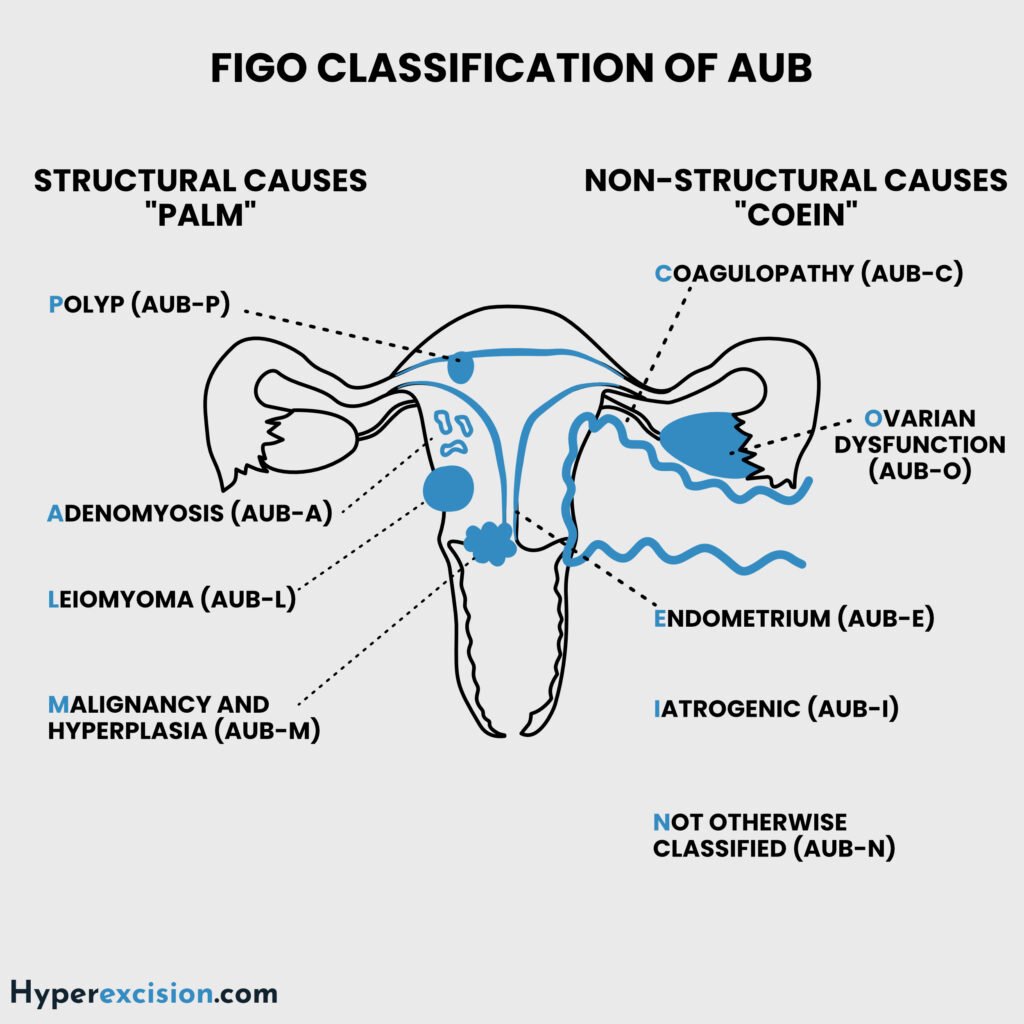
FIGO Classification of Abnormal Uterine Bleeding (PALM-COEIN)
Structural Causes (PALM)
| Acronym | Description |
|---|---|
| P | Polyps (AUB-P) |
| A | Adenomyosis (AUB-A) |
| L | Leiomyoma (AUB-L; submucosal most relevant) |
| M | Malignancy or hyperplasia (AUB-M) |
Non-Structural Causes (COEIN)
| Acronym | Description |
|---|---|
| C | Coagulopathy (AUB-C; e.g. von Willebrand disease) |
| O | Ovulatory dysfunction (AUB-O; commonly anovulation) |
| E | Endometrial (AUB-E; primary disorder) |
| I | Iatrogenic (AUB-I; e.g. anticoagulants, IUD) |
| N | Not classified e.g. niche, arteriovenous malformation |
Q7. Which questions are important to ask in her history?
Reveal answer
Questions on conception
Has the couple been able to conceive before?
Has either member of the couple conceived before (with another partner)?
Gynaecological history
Cycle length
Duration of bleeding
Post-coital bleeding
Sexual history
When and how often does the couple have sex?
Dyspareunia?
What is his libido? (low libido = androgen insensitivity)
History of STDs or genitourinary infections (PID)
Obstetric history
Miscarriages
Previous pregnancies
Complications
Infertility history
Duration of infertility
Frequency of intercourse
Partner history
Prior treatments
Medical history
History of thyroid disease
History of coagulation disorders
Surgical history
History of prior uterine procedures, e.g. myomectomy, curettage (Asherman syndrome)
Family history
Family history of malignancy (lynch syndrome/BRCA)
Review of systems
Weight loss
Symptoms of anaemia, e.g. fatigue, palpitations
She reports prolonged periods lasting 8–10 days with passage of clots and associated dysmenorrhea. She denies post-coital bleeding but notes intermenstrual spotting occasionally. There is no history of chronic pelvic pain, weight loss, or systemic symptoms.
Q8. What are the normal vs. abnormal parameters for menstrual bleeding?
Reveal answer
Normal vs Abnormal parameters for menstrual bleeding
| Parameter | Normal Range | Nota bene |
|---|---|---|
| Menarche | 10 – 16 years (mean age 13) | Irregular periods from 3 years after menarche |
| Cycle Frequency | 21–35 days | <21 is polymenorrhoea while >35 days is oligomenorrhoea |
| Bleeding Duration | 2–7 days | It can be up to 8 mm if there is hormonal replacement therapy |
| Menstrual blood loss Volume | 5–80 mL/cycle | >80 mL is heavy menstrual bleeding |
| Cycle Regularity | Variation < 9 days | Variation ≥ 9 days is abnormal |
| Endometrial thickness (pre-menopausal) | 6 – 12 mm | Abnormal if the thickness is out of phase or exceeds 16 mm in the secretory phase |
| Endometrial thickness (post-menopausal bleeding) | < 4 mm | Can be up to 8 mm if there is hormonal replacement therapy |
Q9. Which features in the history indicate heavy menstrual bleeding?
Reveal answer
Soaking ≥ 2 pads/hour
Changing pads every 3 hours
More than 20 pads used in 1 cycle
Passing clots > 1 inch
Needs extra pads in the night
Bleeding soils clothing or bedsheets
Flow lasts > 7 days
Signs and symptoms of anaemia
On abdominal examination, there is a firm, irregular pelvic-abdominal mass corresponding to an 18-week gravid uterus. The mass is non-tender, nodular, and moves with the cervix. No ascites is detected.
Q10. How can you tell that an abdominal mass is pelvic in origin?
Reveal answer
The mass is felt by palpating from the xiphoid process inferiorly
It arises from the pelvis and grows cephalad
Size corresponds to uterine size in weeks of gestation
A mass that is of uterine origin moves with the cervix and is continuous with the uterus on bimanual examination
Laboratory results:
| Test | Result | Reference Range |
|---|---|---|
| Hemoglobin | 8.9 g/dL | 12–16 g/dL |
| MCV | 74 fL | 80–96 fL |
| Serum Ferritin | 9 ng/mL | 15–150 ng/mL |
| β-hCG | Negative | — |
| FSH | 7 IU/L | 3–10 IU/L (follicular phase) |
| LH | 6 IU/L | 2–12 IU/L |
| TSH | 2.1 µIU/mL | 0.4–4.0 µIU/mL |
| Prolactin | 14 ng/mL | 5–25 ng/mL |
Q11. Interpret these laboratory results
Reveal answer
Low hemoglobin, MCV and serum ferritin indicate iron deficiency anaemia
Negative B-hCG excludes pregnancy
Normal FSH, LH, TSH and prolactin mean normal ovulatory function
Q12. What other investigations are important in this case?
Reveal answer
Transvaginal ultrasound (TVUS): to identify structural lesions and measure endometrial thickness
Coagulation panel: to identify coagulopathy
Hysteroscopy: to identify focal lesions, assess uterine cavity and tubal patency for infertility
Endometrial biopsy is indicated in high-risk patients (> 45 years old and unopposed estrogen exposure)
Further infertility workup may include the partner’s semen for analysis
Q13. What is the most likely diagnosis?
Reveal answer
The diagnosis is Uterine leiomyoma (fibroids)
Heavy menstrual bleeding (the most common symptom)
Q14. What are the risk factors for fibroids?
Reveal answer
Reproductive years (30 – 40 years)
African
Family history (2X risk with primary relative)
Overweight/obesity
Early menarche
Fibroids are estrogen-dependent tumours.
They promote their growth by creating a hyperestrogenic environment (have more estrogen receptors, convert less estradiol to estrone and contain more aromatase)
They usually regress after menopause
Q15. What are the risk-reducing factors for fibroids?
Q16. What are the types of fibroids by location?
Reveal answer
| Type of Fibroid | Clinical association |
|---|---|
| Submucosal (FIGO 0–2) | Heavy menstrual bleeding, infertility, recurrent miscarriage, irone deficiency anaemia |
| Intramural (FIGO 3–5) | Most common type. Associated with heavy menstrual bleeding, pelvic pain, infertility |
| Subserosal (FIGO 6–7) | Pelvic pressure, frequency and urgency, constipation, minimal effect on fertility, rarely associated with abnormal bleeding, can undergo torsion if pedunculated |
| Cervical fibroids (rare) | Dyspareunia, dysuria, urinary obstruction, obstructed labour |
| Intraligamentary (Broad ligament fibroids) | Hydronephrosis |
| Parasitic fibroids | Rare. Can mimic adnexal or abdominal masses. |
| Pedunculated fibroids | Torsion |

Q17. How do fibroids cause heavy menstrual bleeding (HMB)?
Reveal answer
| Pathology | Description |
|---|---|
| Anatomical effects | Increased endometrial surface-area and dilatation of venous channels (venous ectasia or venous lakes) |
| Vascular changes | Upregulation of angiogenic mediators e.g. VEGF, eNOS and related pathways |
| Disturbed myometrial contractility | Abnormal contraction prevents vasoconstriction of spiral arteries which impairs hemostasis |
Q18. How do fibroids cause subfertility?
Reveal answer
| Pathology | Description |
|---|---|
| Anatomical effects | Submucosal fibroids may physically prevent embryo implantation or block tubal ostia |
| Junctional-zone dysfunction | Intramural or submucosal fibroids disrupt the junctional zone (which generates peristalsis) causing impaired sperm transport and embryo positioning |
Q19. What options exist for the medical treatment of uterine bleeding?
Reveal answer
| Medical treatment | Description |
|---|---|
| NSAIDs and tranexamic acid/mefenamic acid | Usually prescribed for 5 days. Controls bleeding in 60% of patients. NSAIDs reduce blood loss by 30% and tranexamic acid reduces blood loss by 40 – 50% |
| High-dose progestin | Usually norethisterone which is prescribed for 10 days. Stabilizes the endometrium |
| Combined oral contraceptive pills | 80% of patients responds to COCs. Preferred for AUB-O |
| Hormonal IUD (Mirena/LNG-IUD) | Most effective hormonal agent. Reduces bleeding by 95%. Preferred for AUB-A. Sufficient distortion of the uterus by fibroids can dislodge the intrauterine device |
| Iron supplementation | For anaemia |
Balloon tamponade can be used for acute intractable bleeding
Q20. What is the role of GnRH agonists/antagonists in the treatment of fibroids?
Reveal answer
Pre-operative therapy to shrink fibroids (facilitates myomectomy or hysterectomy)
Fibroids refractory to combined oral contraceptives
They suppress FSH/LH, which causes a hypoestrogenic state similar to menopause
Agonists include leuprolide, nafarelin, goserelin, and buserelin
Antagonists include relugolix and elagolix
ACOG recommendsa duration of < 6 months without add-back therapy due to side effects of hypoestrogenism (osteoporosis, hot flashes, etc.)
Antagonists are better tolerated for long-term use. Used with add-back therapy (low-dose oestrogen and progestin) to reduce side effects
Q21. What is the definitive treatment for this patient?
Reveal answer
Myomectomy
Myomectomy is preferred if fertility is the main issue of concern. It can be performed laparoscopic, hysteroscopically or open. Future deliveries will need an elective caesarean delivery
Hysterectomy may be performed if fertility is not desired.
Uterine artery embolisation is a minimally invasive option. It is associated with post-embolisation syndrome (pain due to necrosis and cytokine release)
Q22. What degenerative changes can fibroids undergo?
Reveal answer
| Type of Degeneration | Description |
|---|---|
| Hyaline degeneration (most common, ~60%) | Most common degeneration and clinically insignificant. Smooth muscle fibers are replaced by homogeneous eosinophilic hyaline material. |
| Cystic degeneration (~4%) | Mainly post-menopausal due to necrosis. Liquefaction of hyaline areas → cystic spaces filled with serous fluid. May mimic ovarian cysts or molar pregnancy in women of childbearing age |
| Red (carneous) degeneration | Acute hemorrhagic infarction of fibroid, often during pregnancy (2° to venous thrombosis or arterial compromise). An important cause of acute abdomen in pregnancy |
| Myxoid degeneration | Accumulation of myxoid (gelatinous, mucoid) material within fibroid. |
| Fatty (lipomatous) degeneration | Deposition of fat within fibroid tissue. |
| Calcific (dystrophic) degeneration | Calcium deposits within fibroid (especially post-menopausal). |
| Suppurative degeneration | Rare. Infection of necrotic fibroid tissue (especially after uterine instrumentation or postpartum). |
| Sarcomatous degeneration | Malignant transformation. |
2/3 of fibroids undergo degeneration
Q23. What are the obstetric complications of fibroids?
Reveal answer
Miscarriage
Preterm delivery
Fetal malpresentation
Fetal growth restriction
Abnormal placentation
Obstruction of labour