A 42-year-old woman presents to the outpatient clinic with a 6-month history of joint pain and stiffness.
Q1. What are the differentials for joint pain?
| Classification | Differentials |
|---|---|
| Trauma | Sprain, strain, fracture, dislocation, tendon or ligament, or meniscal tear, tendonitis |
| Infectious (septic arthritis) | Gonococcal, non-gonococcal, Lyme disease, viral, tuberculosis, and fungal causes |
| Crystal arthropathies | Gout, pseudogout |
| Degenerative | Osteoarthritis |
| Malignancy | Solid tumor metastasis, lymphoma, leukemia, osteosarcoma, osteochondroma |
| Inflammatory | Rheumatoid arthritis, Reactive arthritis, psoriatic arthritis, systemic lupus erythematosus, Sjogren syndrome, ankylosing spondylitis, acute rheumatic fever |
Q2. Define the following terms: arthralgia, arthritis, and synovitiser
| Term | Definition |
|---|---|
| Arthralgia | Pain that is at the joint or referred to the joint. |
| Arthrtitis | Inflammation of the joint structure (the pathological process) |
| Synovitis | Inflammation of the synovial fluid. Any systemic inflammatory disease can cause synovitis (manifesting as arthralgia) since synovial fluid is an ultrafiltrate of the serum |
Q3. What are the likely causes of arthritis according to age and sex?
| Male | Female | |
|---|---|---|
| Young | Reactive arthritis, ankylosing spondylitis, psoriatic arthropathy, enteropathic arthropathy | SLE, Rheumatoid arthritis, psoriatic arthropathy, enteropathic arthropathy |
| Middle age | Gout, generalised osteoarthritis, polymyagia rheumatic | Rheumatoid arthritis, sicca syndrome, generalised osteoarthritis, polymyagia rheumatic |
| Elderly | Polymyalgia rheumatic |
Q4. What questions are important to ask in the history of a patient with joint pain?
Systemic symptoms: Does the patient have other systemic symptoms, e.g., fever, chills, fatigue, rash, etc?
Location: Which joint is sore? Is it one, some, or multiple? Is it symmetrical or asymmetrical:
Inflammation: Is the joint inflamed? (erythematous, warm, swollen)
Chronicity: Is it a recent onset (acute) or an insidious onset (chronic)? Does the pain come and go, or is it constant? What time of day is the pain worse?
Evidence of trauma: Does the patient have any factors in their history that point to trauma?
SLICE
The pain initially started in the small joints of her hands and has progressively worsened. She describes morning stiffness lasting more than an hour, which improves as the day progresses. She reports swelling, warmth, and decreased grip strength. She also reports experiencing general fatigue, a low-grade fever, and weight loss over the past few months.
Q5. What is the significance of prolonged joint stiffness that is worse in the morning, then improves as the day progresses?
Suggests inflammatory joint pain
Due to cytokine surge overnight, immobility (inflammatory synovial fluid accumulates and thickens), and cortisol nadir at around 2 – 4 am (reduced anti-inflammatory protection)
Degenerative joint disease (osteoarthritis), on the other hand, has brief morning stiffness (< 30 minutes) that worsens with use
Upon examination, both hands exhibit symmetrical swelling and tenderness in the MCP and PIP joints, accompanied by mild ulnar deviation. The wrists are also tender and slightly limited in movement. There are no nodules seen over the extensor surfaces. Cardiovascular, respiratory, and neurological exams are unremarkable.
These are her laboratory results:
| Test | Result | Reference Range |
|---|---|---|
| Hemoglobin | 10.5 g/dL | 12–16 g/dL |
| WBC count | 6.2 × 10⁹/L | 4–11 × 10⁹/L |
| Platelet count | 420 × 10⁹/L | 150–450 × 10⁹/L |
| ESR | 72 mm/hr | <20 mm/hr |
| CRP | 25 mg/L | <5 mg/L |
| Rheumatoid factor (RF) | Positive (120 IU/mL) | <20 IU/mL |
| Anti-CCP antibodies | Positive (high titer) | Negative |
| ANA | Negative | Negative |
| Serum creatinine | 0.9 mg/dL | 0.6–1.2 mg/dL |
| Liver function tests | Normal | Normal |
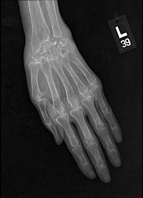
Her X-ray is shown below:
Q6. Interpret the lab findings
Low haemoglobin suggests anaemia of chronic disease
Elevated platelet count suggests inflammation-related thrombocytosis
Elevated ESR and CRP suggest inflammation
Positive rheumatoid factor and anti-CCP suggest rheumatoid arthritis
Negative ANA helps to rule out SLE
Normal serum creatinine suggests preserved kidney function
Normal liver function tests mean no hepatic involvement
Q7. What is the most likely diagnosis?
Joint pain for 6 months
Systemic symptoms (fatigue, fever, and weight loss)
Affects symmetric small-joint polyarthritis
Morning stiffness > 1 hour
Systemic symptoms
Positive Rheumatoid Factor and anti-CCP antibodies
Elevated CRP and ESR
A positive rheumatoid factor is seen in 70% of cases of rheumatoid arthritis. High titres are associated with severe disease, erosions, and extra-articular disease
Anti-CCP antibodies are highly specific for rheumatoid arthritis (98%) and moderately sensitive (70-80%)
Summary of diagnostic criteria for RA (≥ 6 points required)
Q8. What are the diagnostic criteria for this condition?
2010 ACR/EULAR classification criteria:
- Joint involvement (number and site of joints)Serology for rheumatoid factor or anti-CCP (low positive vs high positive)Elevated acute-phase reactants (ESR, CRP)Duration of symptoms (at least 6 weeks)
Q9. What is the pathophysiology of this condition?
Genetic susceptibility
Strong association with HLA-DR4 and HLA-DR1 alleles, which present self-antigens (citrullinated peptides) to T-cells, promoting autoimmunity
Environmental triggers
Cigarette smoking induces citrullination of proteins in the lungs
Other triggers include periodontal infections (Porphyromona gingivalis), viral infections, and microbiome changes
Loss of tolerance
The immune system fails to recognize these citrullinated proteins as “self.” This leads to the production of anti-citrullinated protein antibodies (ACPAs), measured clinically as anti-CCP
Inflammation
Citrullinated peptides are recognized as antigens, and autoantibodies are formed against them → autoantibodies form immune complexes, which are deposited in joints → complement activation → inflammation occurs
Rheumatoid factor (RF) is an IgM antibody against the Fc portion of IgG
Anti-citrullinated protein antibodies (Anti-CCP) are highly specific for rheumatoid arthritis
Immune cells infiltrate the synovium, promoting inflammation and joint damage
Systemic symptoms are due to circulating cytokines (IL-6 and TNF-α)
Joint changes
Chronic synovitis → hyperplasia of synovial tissue (pannus – proliferating fibroblast-like synoviocytes, inflammatory cells and granulation tissue) → pannus invades cartilage and bone → erosion and joint destruction
Osteoclasts are activated via RANK/RANKL → marginal bone erosion and joint deformities
Q10. What are the extra-articular features of this condition?
Extra-articular manifestations of RA
| Classification | Extraarticular manifestations |
|---|---|
| Nodules | Elbow, lungs, cardiac, CNS, lymphadenopathy, vasculitis |
| Lungs | Pleural disease, interstitial fibrosis, bronchiolitis obliterans, organizing pneumonia |
| Cardiac | Ischemic heart disease, pericarditis, pericardial effusion, carpal tunnel syndrome, peripheral neuropathy, splenomegaly (including Felty’s syndrome) |
| Musculoskeletal | Carpal tunnel syndrome, peripheral neuropathy, osteoporosis |
| Ophthalmologic | Episcleritis, scleritis, scleromalacia, keratoconjuncitivitis sicca |
| Haematologic | Anaemia, splenomegaly (including Felty’s syndrome), amyloidosis |
Q11. What physical exam findings are characteristic of this condition?
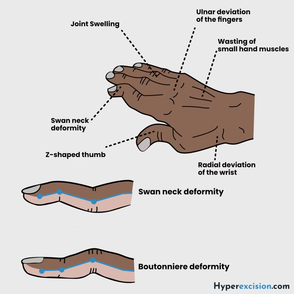
Symmetrically swollen MCP, PIP, wrist or MTP joints
Ulnar deviation and subluxation of wrist and fingers
Boutonniere and swan-neck deformitis of fingers
Rheumatoid nodules on bony prominences
Z-deformity of thumbs
Foot changes (similar to hand changes)
Atlanto-axial joint sublaxation (rare)
Baker’s cyst
Q12. What radiographic features are characteristic of this condition?
Soft tissue swelling
Juxta-articular osteopaenia
Reduced joint spacing
Bony erosions
Subluxation
Complete carpal destruction (late)
Q13. What is the pharmacological treatment of this condition?
| Category | Description |
|---|---|
| NSAIDs | For symptomatic relief |
| Intra-articular or intramuscular steroids | For acute exacerbation or as a bridge to DMARDs |
| Disease-modifying antirheumatic drugs (DMARDs) | First-line agents. Started within 3 months of persistent symptoms. Takes 6 – 12 weeks for symptomatic benefit. Different DMARDs can be combined. Early prescription improves long-term outcomes |
| Biological agents | Early prescription of biological agents improves long-term outcomes. These include TNF-α inhibitors, B-cell depletion, IL-1 and IL-6 inhibition, and inhibition of T-cell co-stimulation |
DMARDs include methotrexate, sulfasalazine, hydroxychloroquine, and leflunomide
Biological agents include TNF-A inhibitors (infliximab, etanercept, adalimumab), B-cell depletion (rituximab), IL-1 and IL-6 inhibition (tocilizumab), and inhibition of T-cell co-stimulation (abatacept)
Surgery may include synovectomy or arthroplasties
Q14. What laboratory investigations does this patient require before starting treatment with DMARDs?
Liver function tests
Hepatitis serology
Pregnancy test
These tests are important to get before prescribing methotrexate
Q15. What are the side effects of DMARDs?
Side effects of DMARDs
| Category | Side effects |
|---|---|
| General | Immunosuppression, pancytopaenia (especially if combined with methotrexate |
| Methotrexate | Pneumonitis, oral ulcers, hepatotoxicity, teratogenic |
| Sulfasalazine | Rash, reduced sperm count, oral ulcers, gastrointestinal upset |
| Leflunomide | Teratogenic, oral ulcers, hypertension, hepatotoxicity |
| Hydroxychloroquine | Retinopathy |
Q16. What are the side effects of biological agents?
Serious infection
Reactivation of TB and hepatitis B
Worsening heart failure
Hypersensitivity
Injection-site reactions and blood disorders
Evolution of ANA and reversible SLE-type illness
Q17. Define seronegative spondyloarthropathies. What are their diagnostic hallmarks?
Seronegative spondyloarthropathies are immune-mediated inflammatory diseases that tend to affect the axial skeletal lack rheumatoid factor, and anti-CCP antibodies
PAIR: Psoriatic arthritis, ankylosing spondyloarthritis, IBD-associated (enteropathic arthritis), reactive arthritis
Diagnostic hallmarks of seronegative spondyloarthropathies
| Features | Description |
|---|---|
| Seronegativity | Rheumatoid factor and anti-CCP negative |
| Genetic association | HLA-B27 |
| Main sites involved | Axial arthritis (axial skeleton) – spine and sacroiliac joints |
| Other sites involved | Entheses (achilles tendonitis, plantar fasciitis), dactylitis, peripheral joints |
| Extra-articular manifestations | Anterior uveitis, psoriatic rash, oral ulcers, aortic valve incompetence, and inflammatory bowel disease |
Q18. What is the difference between osteoarthritis, rheumatoid arthritis, psoriatic arthritis, reactive arthritis, ankylosing spondylitis, and septic arthritis?
| Osteoarthritis | Rheumatoid arthritis | Psoriatic arthritis | Reactive arthritis | Ankylosing spondylitis | Septic arthritis | |
|---|---|---|---|---|---|---|
| Patient history | Old and overweight | Young woman | Middle aged | Young person, history of infection | Young man, morning stiffness | Sexually active, joint prostheses, history of osteomyelitis |
| Systemic symptoms | None | Systemic symptoms are present + rheumatoid nodules, and glomerulonephritis | Systemic symptoms are present, + psoriasis | Systemic symptoms are present, + urethritis and ocular involvement | Systemic symptoms are present | Sepsis |
| Location | Weight-bearing joints and fingers | Small joints, spares the DIPs | Fingers, including DIPs | Knee, ankles, feet | Lumbosacral spine, pelvis | Knee (most common) |
| Inflammation | No | Yes | Yes | Yes | Yes | Yes (severe) |
| Chronicity | Long-term, worse at night | Long-term, worse in the morning | Long-term worse in the morning | Weeks, worse in the morning | Long-term worse in the morning | Acute |
| Evidence of trauma | None | None | None | None | None | Possible |
| Investigations | Joint space narrowing and spurs on X-ray | Rheumatoid factors, anti-CCP | None; HLA-B27 positive | None; HLA-27 positive | Bamboo spine on X-ray, HLA-B27 positive | Purulent synovial fluid; leukocytosis |