Overview
Acute coronary syndrome (ACS) is a spectrum of clinical presentation ranging from ST-segment elevation myocardial infarction (STEMI), non-ST-segment elevation myocardial infarction (NSTEMI), and unstable angina. It is caused by rupture of an atherosclerotic plaque and partial or complete occlusion of a coronary artery.
It peaks between 60 and 70 years, and affects men more than women.
Definition of terms
| Term | Definition |
|---|---|
| Ischemic heart disease (IHD) | An imbalance between myocardial perfusion and demand for oxygen |
| Myocardial ischemia | Reduced blood supply +/- death of myocardial cells |
| Acute myocardial infarction | Death of myocardial cells with the release of cardiac enzymes |
| Silent myocardial infarction | An asymptomatic infarction occurs in 20% of cases. Common in the elderly and in patients with diabetes |
Types of ACS
| Type of ACS | Description | Findings |
|---|---|---|
| Non-MI Unstable angina | Angina of increasing frequency or intensity, or occurring at rest | No elevated enzymes |
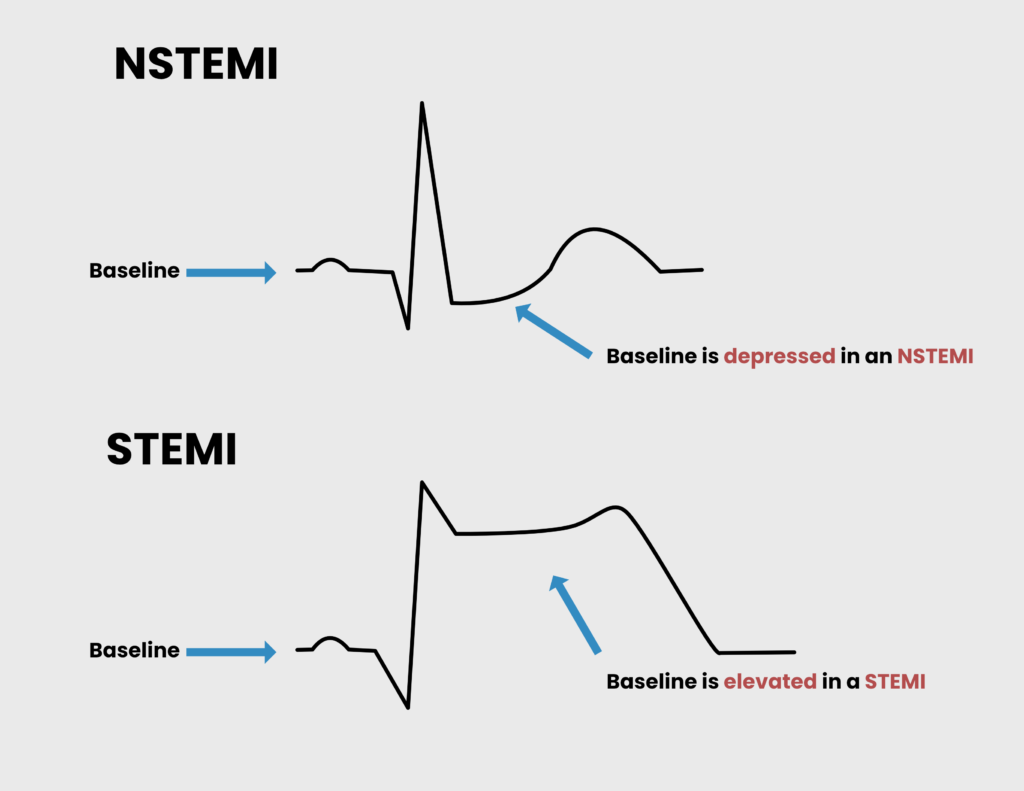
| NSTEMI | ACS without ST-segment elevation | ST depression (Ischemia) + elevated enzymes |
| STEMI | ACS with ST-segment elevation or new-onset LBBB | ST elevation (Infarction) + elevated enzymes |
| Stable Angina | Unstable Angina | NSTEMI | STEMI | |
|---|---|---|---|---|
| Pain | Exertion | Rest | Rest | Rest |
| Relief | Rest + Nitrate | No | No | No |
| Biomarkers | No | No | Yes | Yes |
| ST changes | No | No | No | Yes |
| Stenosis | 70% | 90% | 90% | 100% |
- Risk factors for ACS
- Modifiable
- Cholesterol level (Target LDL < 100; <70 if diabetic)
- Smoking
- Hypertension
- Diabetes Mellitus
- Obesity
- Sedentary lifestyle
- Unhealthy diet – saturated fats, trans fats, salt, and refined sugars
- Excess alcohol consumption
- Psychosocial stress
- Non-modifiable
- Age > 60 years
- Male gender (and post-menopausal women)
- Family History of Myocardial Infarction
- Modifiable
- Non-coronary atherosclerotic causes of Myocardial infarction
- Vasculitis (Polyarteritis Nodosa, Kawasaki disease)
- Cocaine use
- Thrombophilia (Antithrombin III deficiency, Polycythemia vera)
- Aortic dissection
- Iatrogenic (after revascularization procedure)
- Pathophysiology
- Disruption of an atheromatous plaque in a coronary artery → subendothelial collagen and thrombogenic material (Tissue thromboplastin) are exposed → Platelets adhere to exposed material → occlusion of the artery by thrombus → Ischemia
- Irreversible myocardial injury
- Mitochondrial dysfunction: ischemia alters the permeability of the mitochondrial membrane, causing swelling, rupture of the outer membrane, and release of pro-apoptotic proteins
- Formation of superoxide free radicals: damages myocytes by altering membrane proteins and phospholipids
- Acute inflammation with neutrophil infiltration: reduces blood flow to ischemic tissue by occluding the capillary lumen (non-reflow phenomenon). Also releases proteolytic enzymes and reactive oxygen species.
- Activation of the complement system, which further damages the endothelium
- Ischemia-reperfusion injury
- Spontaneous reperfusion or reperfusion following PCI/tPA → newly returning blood carries reactive oxygen species and ions → damage to the cell membrane → influx of Ca2+ → contraction of myofibrils → cytoskeletal damage and contraction band necrosis
- Signs and symptoms
- Chest pain
- Severe
- Retrosternal/epigastric
- Crushing, burning, or discomfort
- Radiates to the neck and down the inner part of the left arm
- Last 20 minutes – 7 hours
- Occurs at rest
- Pallor
- Diaphoresis
- Dyspnoea
- Nausea and vomiting
- Palpitations
- Hypotension
- Syncope or presyncope
- Fatigue
- Chest pain
- Differentials
- Stable angina
- Pericarditis
- Myocarditis
- Aortic dissection
- Takotsuba cardiomyopathy
- Arrhythmia
- Pneumothorax
- Pulmonary embolism
- Pneumonia
- Pleurisy
- Gastroesophageal reflux disease
- Peptic ulcer disease or gastritis
- Pancreatitis
- Musculoskeletal pain (chostochondritis)
- Investigations
- Electrocardiography to determine whether it is NSTEMI or STEMI
- Hyperacute T waves: often the first sign of myocardial infarction, but persists for a few minutes
- Biphasic or inverted T-waves (Wellen’s syndrome): areas of ischemia at the periphery of the infarct. Occurs at rest. Inversion can last for days to months
- Elevated ST segment: injured myocardial cells surrounding the area of necrosis
- New Pathological Q waves: areas of coagulation necrosis. Develops after several hours to days and persists indefinitely.
- Cardiac enzymes to determine whether there is an infarction or not
- Serial testing is required to rule out infarction since neither CK-MB nor Troponins consistently appear in blood within 6 hours of ischemia
- CK-MB: Good for assessing reinfarction. Appears in the first 4-12 hours. Corrects within 3 days.
- Cardiac Troponins: Highly sensitive (96%). Appears within 3-12 hours. Corrects within 7-12 days.
- Echocardiography to look for regional wall abnormalities
- Chest X-ray to look for cardiomegaly, pulmonary oedema, or a widened mediastinum
- Other Labs: Complete blood count, U/E/C, Random Blood Sugar, and Lipids
- Electrocardiography to determine whether it is NSTEMI or STEMI
- Emergency Treatment of ACS with presumptive EKG diagnosis while awaiting cardiac enzymes
- Cardio-pulmonary resuscitation (CPR)
- Morphine to alleviate severe pain and anxiety
- 100% Oxygen to improve saturation in patients with hypoxia (SpO2 < 94%) or respiratory distress
- Sublingual or intravenous nitrates to induce vasodilation and reverse vasospasm
- Aspirin to reduce clot propagation
- Beta-blocker or Calcium Channel Blocker to reduce myocardial oxygen demand
Bottomline treatment of ACS
| Treatment | When to use it | Treatment goal |
|---|---|---|
| Statin | Any ACS | LDL < 70 and HDL > 40 |
| Beta Blocker | Any ACS | SBP < 140 and DBP < 90 |
| ACEi/ARB | Any ACS | SBP < 140 and DBP < 90 |
| Aspirin | Any ACS | No goal |
| Clopidogrel | Aspirin allergy or drug-eluting stent | |
| tPA | STEMI or positive stress test when PCI or transport is unavailable | |
| Fondaparinux | NSTEMI patients who are not at a high risk of bleeding and who are not having angiography immediately | |
| Unfractionated heparin | NSTEMI if immediate angiography is planned or creatinine is > 265 umol/L. ST elevation or positive stress test when tPA is contraindicated. | |
| Angioplasty (PCI) | STEMI or positive stress test, 1 or 2 vessel disease | |
| CABG | STEMI or positive stress test, Left-mainstem or 3 vessel disease |
Gross morphology of myocardial infarction
| Time | Gross findings |
|---|---|
| 0 – 4 hours | None |
| 4 – 12 hours | Dark mottling; infarcted tissue |
| 12 – 24 hours | Dark mottling; infarcted tissue |
| 1 – 3 days | Hyperemia |
| 3 – 14 days | Hyperemic border with central yellow-brown pallor and softening |
| 2 weeks to months | Grey-white scar, recanalized artery |
Histology of Acute Myocardial Infarction
| Time | Histological findings |
|---|---|
| 0-4 hours | None |
| 4 – 12 hours | Early coagulative necrosis |
| 12 – 24 hours | Coagulative necrosis, neutrophil migration |
| 1-3 days | Coagulative necrosis, neutrophil migration |
| 3-14 days | Macrophages, granulation tissue |
| 2 weeks to months | Fibrosis |
STEMI vs NSTEMI
| EKG | Pathology | Treatment |
|---|---|---|
| NSTEMI | Occlusion that causes subendocardial ischemia | Heparin can be used to prevent clot extension |
| STEMI | More severe occlusion that causes transmural ischemia | Fibrinolytics (tPA) can be used to break the clot down |
- Definition of an ST elevation
- New ST elevation at the J-point in two continuous leads
- Cut-off points
- ≥ 0.2 mV in men or ≥ 0.15 mV in women in leads V2-V3
- ≥ 0.1 mV in other leads
- STEMI criteria
- Clinical symptoms consistent with ACS with persistent EKG features in ≥ 2 contiguous leads of
- 2.5 mm (≥ 2.5 small squares) ST elevation in leads V2-V3 in men under 40 years
- 2.0 mm (≥ 2.0 small squares) ST elevation in leads V2-V3 in men over 40 years
- 1.5 mm ST elevation in V2-V3 in women
- 1mm ST elevation in other leads
- New left bundle branch block (LBBB)
- Clinical symptoms consistent with ACS with persistent EKG features in ≥ 2 contiguous leads of
- Similarities between STEMI and NSTEMI
- Both cause cardiac enzyme elevation
- Both are life-threatening emergencies that require treatment
- Both can be classified as anterior, lateral, inferior, anteroseptal, etc.
- Differences between STEMI and NSTEMI
- STEMI infarcts have a higher early mortality rate compared to NSTEMI
- NSTEMI infarcts have an increased risk of reinfarction and Sudden Cardiac Death post-MI
EKG changes and coronary territories
| Location | ECG changes | Coronary artery |
|---|---|---|
| Anteroseptal | V1-V4 | Left anterior descending |
| Inferior | II, III, aVF | Right coronary |
| Anterolateral | V1-6, I, aVL | Proximal left anterior descending |
| Lateral | I, aVL +/- V5-6 | Left circumflex |
| Posterior | Changes in V1-3. Reciprocal changes of STEMI are typically seen: horizontal ST depression, tall, broad R waves, upright T waves, dominant R wave in V2. Posterior infarction is confirmed by ST elevation and Q waves in posterior leads (V7-9) | Usually, the left circumflex, also, the right coronary |
Treatment of Unstable Angina or NSTEMI
- Definitive treatment
- Catheterization for Angiography and potentially Percutaneous Coronary Intervention (PCI)
- Administer Clopidogrel and GP IIb/IIIa inhibitor before PCI – eptifibatide or tirofiban
- Coronary Artery Bypass Graft (CABG)
- Heparin to prevent clot extension
- Catheterization for Angiography and potentially Percutaneous Coronary Intervention (PCI)
- Indications for catheterization for Angiography and Percutaneous Intervention (PCI) in NSTEMI
- Indications for Coronary Artery Bypass Graft (CABG) in NSTEMI
- 12-hour onset
- PCI has failed
Treatment of STEMI
- Definitive treatment
- Tissue plasminogen activator (tPA – alteplase or reteplase) within 3 hours to dissolve the clot and rescue reversibly damaged myocytes
- Admit once recanalization is successful
- tPA should be followed up with either PCI or CABG within 24 hours
- Catheterization for angiography and percutaneous coronary intervention (PCI)
- Administer Clopidogrel and GP IIb/IIIa inhibitor before PCI – eptifibatide or tirofiban
- Rescue PCI can be done if tPA is unsuccessful or contraindicated
- Coronary artery bypass graft (CABG)
- Tissue plasminogen activator (tPA – alteplase or reteplase) within 3 hours to dissolve the clot and rescue reversibly damaged myocytes
Inpatient Treatment of Myocardial Infarction
- Investigations
- Echocardiogram to check for LV function and the geographical damage done to the heart
- Left ventricular ejection fraction is the most significant prognostic factor for survival following myocardial infarction
- Serial Cardiac enzymes to detect reinfarction
- Echocardiogram to check for LV function and the geographical damage done to the heart
- Treatment
- Frequent monitoring every 3 – 4 hours for cardiovascular and pulmonary symptoms
- Aspirin: decreases mortality 😃
- IV beta-blockers: decrease mortality 😃
- Anticoagulation with Heparin or Fondaparinux (INR 1.5 – 2.5): decreases mortality 😃
- Nitroglycerin: for comfort
- Morphine: for pain
- High-dose Statin: The majority of patients with Myocardial infarction will have hypercholesterolemia
- Strict bed rest for 24-48 hours to prevent the heart rate from rising
- Gradually rehabilitate to activity after 48 hours
Outpatient Treatment of Myocardial Infarction
After discharging the patient, we want to minimize the risk of developing MI down the road. Make sure they are on a very strict medical and lifestyle regimen. Follow-up within 2 weeks of discharge.
- Discharge medications
- Aspirin: for all patients for life; reduces clotting (reduces mortality) 😃
- Beta-Blocker: for all patients for life; reduces Oxygen demand (reduces mortality) 😃
- Clopidogrel: for all patients, for at least 9-12 months; reduces clotting (reduces mortality)😃
- Statins: all patients (reduce mortality) 😃
- Nitrates: In all patients, nitroglycerin > ISDN since they can develop tolerance to ISDN – long-acting (reduce pain)
- ACEi or ARB: for patients with LVEF <40% or symptoms of CHF
- Warfarin: for patients at risk of embolism due to A-fib, CHF
- Counselling and lifestyle management (secondary risk factor reduction)
- Hypertension: managed anyway with Beta-blockers (+/- ACEi or ARB, particularly if the patient has Diabetes mellitus. Can also use Diuretics)
- Diabetes: strict management with diet or insulin therapy. Home BG monitoring. HbA1C check q 3 mos.
- Cholesterol: statin is started in all post-MI pts.
- Smoking cessation
- Appropriate physical activity: walking → cycling → swimming → jogging
- OK to have Sex – Male patients may develop psychogenic erectile dysfunction. Diagnosed clinically. It is safe to discontinue nitrates and prescribe sildenafil.
- Stress testing for PCI/CABG (4-7 days after discharge. Protocol as in stable angina. Usually comes back positive. Workup for PCI/CABG afterward)
- Resume driving at 1 month
- Return to work at 3 months
- ABCDE mnemonic for Post-MI long-term management
Complications of Myocardial Infarction
- Complications of MI
- Arrhythmia: most common complication (90%; any kind of arrhythmia under the sun, commonly bradycardia and AV block)
- Ventricular fibrillation: the most common cause of sudden death
- Contractile dysfunction: The patient slowly starts developing CHF symptoms and a new murmur ( LV or RV dysfunction, hence the need for an echo)
- Mechanical dysfunction: The patient suddenly decompensates and has a new murmur (papillary rupture, septal rupture, free wall rupture, hence the need for an echo)
- Reinfarction: will have rebound CK-MB
- Other complications
- Cardiogenic shock (7%): Survival is greatly improved by revascularization
- Anterior wall rupture: can produce cardiac tamponade
- Posteromedial papillary muscle rupture: seen as acute onset mitral regurgitation or LHF
- Interventricular septum rupture: seen as a new RHF caused by left-right shunting
- Mural thrombus (10%): risk of peripheral embolization and stroke
- Fibrinous pericarditis: During the first week of STEMI. Presents as sub-sternal chest pain relieved by leaning forward and aggravated by leaning backward. Friction rub may be present.
- Dressler syndrome: Occurs after the first week of STEMI due to autoantibodies against pericardial antigens. AKA Autoimmune pericarditis
- Ventricular aneurysm: Occurs late (4-8 weeks) due to fibrosis. Complicated by HFrEF due to a lack of contractile tissue.