A 52-year-old woman presents with a 3-month history of a painless lump in her left breast. Biopsy confirms invasive ductal carcinoma, stage II (T2N1M0). She undergoes breast-conserving therapy (lumpectomy with sentinel lymph node biopsy). Post-operatively, she is referred to the oncology clinic where the multidisciplinary team recommends adjuvant radiotherapy to the left breast and regional lymph nodes. She is scheduled for external beam radiotherapy using modern planning techniques.
Q1. What is radiotherapy?
Radiotherapy, also known as radiation therapy, is the use of ionising radiation to destroy cancer cells
Ionising radiation is high-energy radiation that displaces electrons from atoms and molecules. This can be in the form of electromagnetic waves (photons and gamma rays) and particles (neutrons and protons)
A gamma ray is a photon that is emitted spontaneously by a radioactive substance
Q2. Why is radiotherapy indicated for this patient?
Radiotherapy is indicated after breast-conserving surgery to reduce the risk of local recurrence
It also treats potential microscopic residual disease in the breast and regional lymph nodes
The overall survival is also improved with adjuvant radiotherapy
Q3. What are the main techniques of radiotherapy?
| Technique | Description | Examples |
|---|---|---|
| External beam radiotherapy (EBRT) | This is the most common method. Radiation is delivered from a source outside the body | Conventional 2D radiotherapy, 3D conformal radiotherapy (3D-CRT), Intensity-modulated radiotherapy, volumetric modulated radiotherapy (VMAT), Image-guided radiotherapy (IGRT), 4D radiotherapy, stereotactic radiotherapy (SRS/SBRT), proton beam therapy |
| Internal radiotherapy (Brachytherapy) | A radioactive source is placed inside/next to the tumour. This delivers high local radiation doses with minimal exposure to surrounding tissue. | Intracavitary (cervix, uterus), interstitial (prostate, breast) |
| Systemic radiotherapy | Radioactive isotopes are administered systemically either through the oral or intravenous route | Radioactive iodine (I-131 for thyroid cancer), strontium-89 or samarium-153 (for bone metastases), lutetium-177 dotatate (for neuroendocrine tumours) |
Q4. What is the meaning of fractionation of radiotherapy?
Fractionation refers to dividing a treatment over a period of time
Fractionation results in shrinking of the tumour mass over time, making the inner, previously anoxic cells more sensitive to radiation
Q5. What is the mechanism of action of radiotherapy?
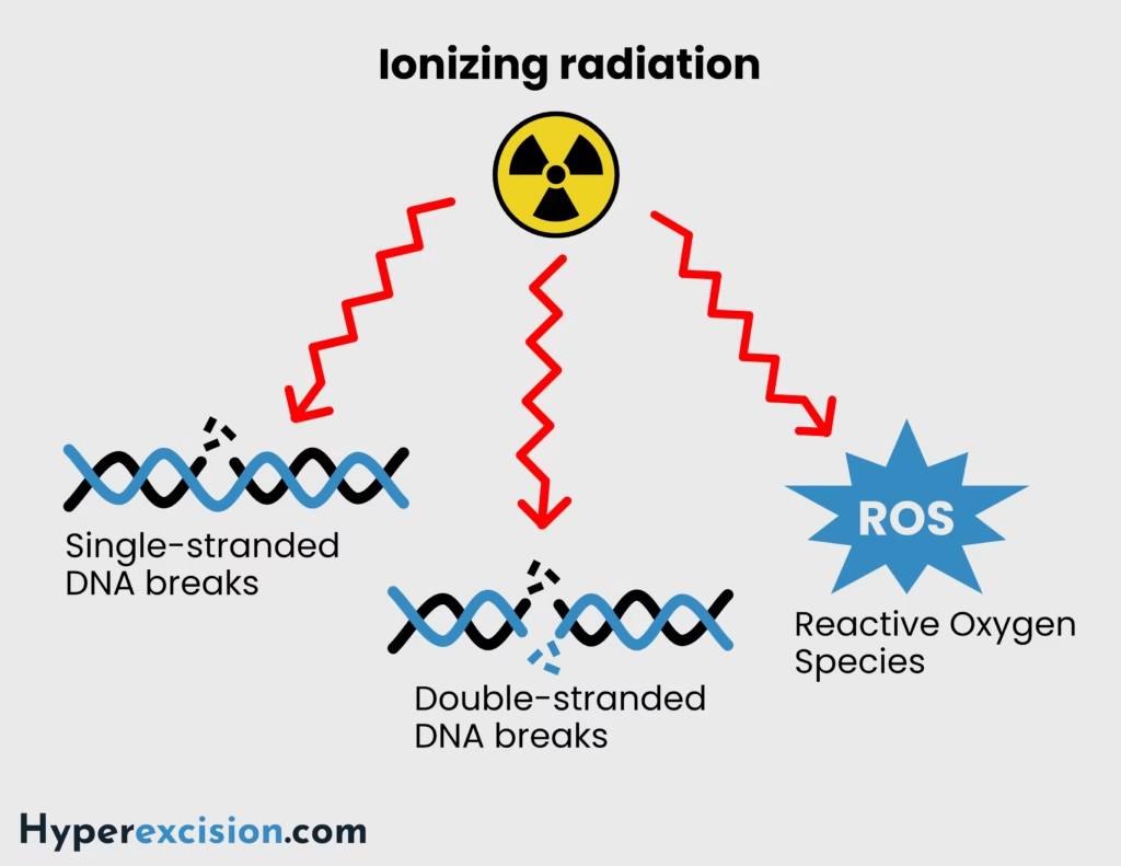
Ionising radiation damages DNA and cellular structures both directly and indirectly
Direct effect is through inducing single and double-stranded breaks in DNA
Indirect effects are through generating reactive oxygen species (ROS), which damage DNA, proteins and membranes
The effect of radiotherapy is based on the 5 R’s (radiobiology of radiotherapy):
Repair: cancer cells have defective DNA repair mechanisms, making them more sensitive to ionising radiation
Redistribution: Fractionation allows cells to move from a radio-resistant phase to a radiosensitive phase (S/M/G2). Redistributes partially synchronised cells to the same part of the cell cycle, causing G2/M block
Reoxygenation: The presence of oxygen prevents normal repair of single and double-stranded DNA breaks. Hypoxic cells limit the curability of cancer.
Repopulation: Killing tumour and normal cells allows both of them to repopulate, but normal cells repair DNA breaks faster than tumour cells, particularly because most of the tumour cells will now be re-oxgenated.
Radiosensitivity: Targeting tissues that are more/less radiosensitive
Cells are most sensitive to ionising radiation in the G2/M phase, while they are relatively resistant in the S phase
Cancer cells are rapidly dividing and have defective DNA repair mechanisms. This makes them more sensitive to ionising radiation than non-cancerous cells.
The outcome is:
Mitotic catastrophe (cells are unable to divide)
Senescence (permanent growth arrest)
Apoptosis or necrosis
Q6. Which tumours are most sensitive to radiotherapy?
| Category | Tumors |
|---|---|
| Most radiosensitive | Lymphoma, germinomas, small cell lung cancer, medulloblastoma, Ewing sarcoma, neuroblastoma, Wilms tumour |
| Moderately radiosensitive | Squamous cell carcinoma, transitional cell carcinoma of the bladder, breast cancer, prostate cancer |
| Radioresistant | Melanoma, renal cell carcinoma, sarcoma (except Ewing sarcoma), glioblastoma multiforme |
Q7. What are the clinical applications of radiation therapy?
| Clinical application | Description | Examples |
|---|---|---|
| Curative therapy | Used for localised cancers where a cure is possible | Head and neck cancer, cervical cancer (via brachytherapy), Hodgkin’s lymphoma, prostate cancer, |
| Adjuvant therapy | Used after surgery to reduce recurrence | Breast cancer, brain tumours |
| Neoadjuvant therapy | Used before surgery to reduce the size of tumours | Rectal cancer, esophageal cancer |
| Palliative therapy | Used for symptom relief in advanced disease | For pain in bone metastases, symptomatic brain metastases, lung cancer, bleeding tumours, and spinal cord compression |
| Prophylactic therapy | Used to prevent metastasis in sanctuary sites | Prophylactic cranial irradiation in small cell lung cancer |
Q8. What are the steps of radiation therapy, including planning?
Consultation and Decision/ preauthorisation and dose prescription
Indication assessed. Clinician fills intent form, consent obtained. Patient file is opened, and dose recorded
Simulation/ positioning and immobilisation
CT/MRI planning scans performed in treatment position
Align the patient to the room coordinate systems
Sedate patients who cannot follow commands, e.g. children
Target Delineation/ contouring
Gross Tumour Volume (GTV)
Clinical Target Volume (CTV)
Planning Target Volume (PTV)
Organs at Risk (OARs) are outlined.
Treatment Planning
Computerised planning system calculates optimal beam arrangement, dose, and shielding
Dose Prescription
Typically measured in Gray (Gy), fractionated (e.g., 2 Gy/day × 25 fractions).
Verification & Quality Assurance
Portal imaging / cone-beam CT to verify accuracy.
Treatment Delivery
Radiation administered as per plan.
Follow-up & Assessment
Monitor response and manage side effects.
In summary: Pateint positioning and immobilisation→ volumetric data acquisition→ image transfer to the TPS→ Target volume delineation→ 3D model→ forward or inverse planning→ dose distribution→ treatment QA→ Treatment delivery
Q9. What are the side effects of radiation therapy?
| System | Examples |
|---|---|
| Constitutional | Fatigue, malaise, anorexia, nausea |
| Skin and connective tissue | Erythema, hyperpigmentation, desquamation, radiation dermatitis, atrophy, telangiectasia, fibrosis, poor wound healing, secondary skin cancer |
| Haematologic | Anaemia, leukopaenia, thrombocytopaenia |
| Gastrointestinal | Mucositis, esophagitis, nausea, vomiting, diarrhoea, abdominal cramps, strictures, ulceration, fistulae, radiation enteritis/proctitis, malabsorptions |
| Respiratory | Radiation pneumonitis, pulmonary fibrosis, restrictive lung disease |
| Cardiovascular | Pericarditis, restrictive cardiomyopathy |
| Neurological | Cerebral edema, somnolence syndrome, radiation myelopathy, cognitive impairment, memory loss, necrosis of brain tissue |
| Endocrine | Hypothyroidism (neck), hypopituitarism (cranial), growth retardiation (skeletal and endocrine), infertility (gonads) |
| Genitourinary | Cystitis, dysuria, frequency, hematuria, contracted bladder, fistula formation, and infertility |
| Ophtlamologic | Conjunctivitis, keratitis, cataracts, retinopathy, optic neuropathy |
| Head and neck | Otitis externa, serous otitis, sensorineural hearing loss, xerostomia, mucositis, dental caries, osteoradionecrosis (mandible) |