Liver cirrhosis is irreversible liver damage that is characterised by diffuse bridging fibrosis and regenerative nodules. It has various causes, all of which lead to inflammation and damage to hepatocytes. Fibrosis causes increased resistance to blood flow in the liver, which leads to portal hypertension.
The overall 5-year survival for cirrhosis is 50%.
Differentials for portal hypertension
| Category | Differentials |
|---|---|
| Pre-hepatic (portal vein) | Portal vein thrombosis, structural abnormality of the portal vein (atresia or stenosis) |
| Intrahepatic (sinusoidal or parenchymal) | Cirrhosis, PBC (even in the abscence of cirrhosis), nodular regenerative hyperplasia, schistosomiasis, massie fatty change, sarcoidosis, infiltrative malignancy, amyloidosis |
| Post-hepatic (hepatic veins and beyond) | Right heart failure, budd-chiari syndrome, constrictive pericarditis |
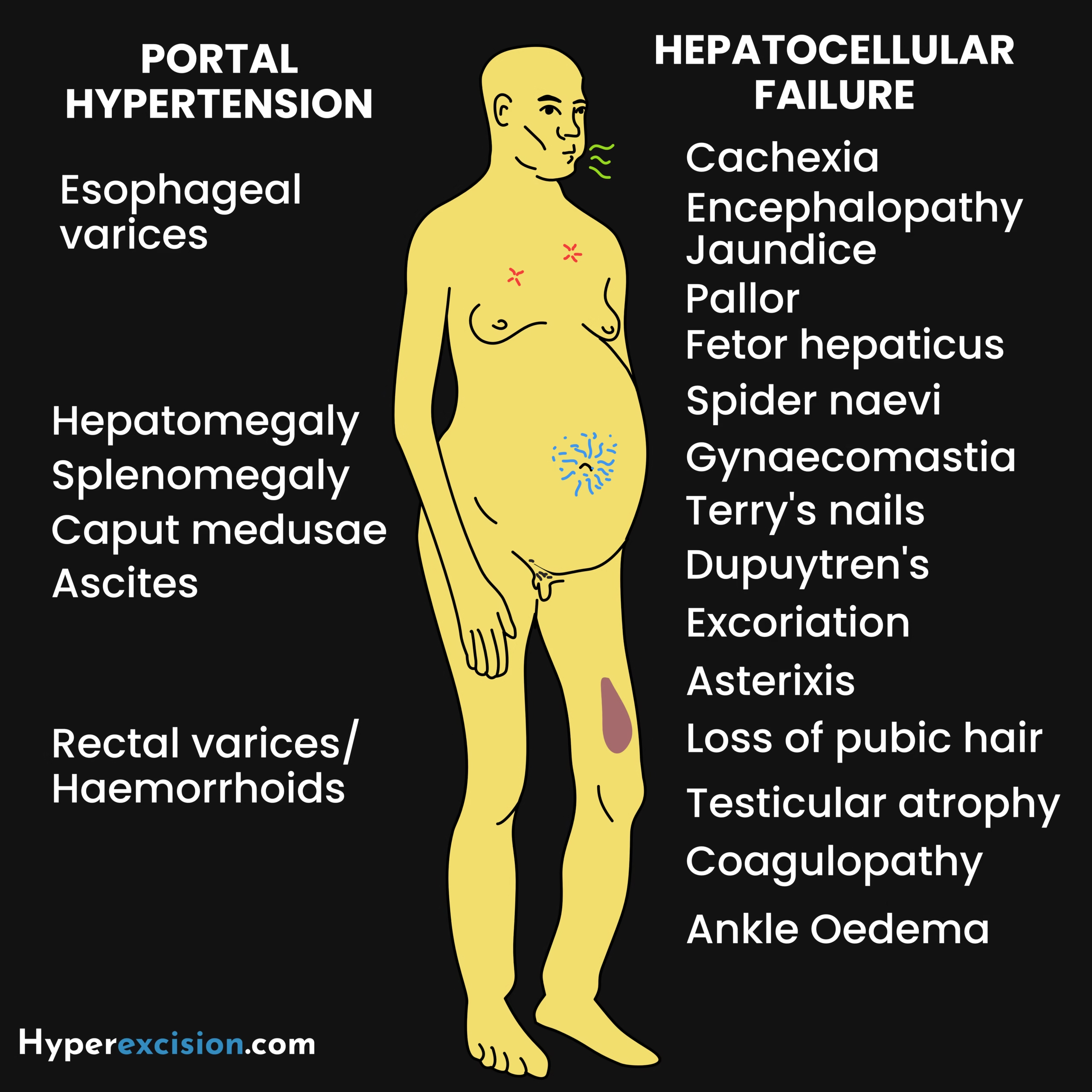
- Causes
- Chronic alcohol abuse (most common cause)
- Non-alcoholic steatohepatitis (NSAH)
- Infectious disease
- Chronic Hepatitis B and Hepatitis C (post-necrotic cirrhosis)
- Schistosomiasis
- Genetic disorders
- Haemochromatosis
- Alpha-1 antitrypsin deficiency
- Wilson’s disease
- Cystic fibrosis
- Vascular disease
- Budd-chiari syndrome
- Autoimmune disease
- Primary biliary cholangitis (PBC)
- Autoimmune hepatitis
- Primary sclerosing cholangitis
- Drugs
- Amiodarone
- Methyldopa
- Methotrexate
- Sodium valproate
- Cryptogenic cirrhosis
- Pathophysiology
- Chronic liver injury → activation of hepatic stellate cells → fibrosis → impaired sinusoidal blood flow → portal hypertension and reduced hepatocellular function
- Splanchnic vasodilatation and portal hypertension → portosystemic collaterals, splenomegaly, and ascites
- Loss of functional hepatocytes:
- Hypoalbuminaemia
- Coagulopathy
- Reduced detoxification
- Cholestasis
- Impaired immune surveilance → increased susceptibility to infections
- Signs and symptoms
- Cachexia
- Jaundice
- Hepatomegaly
- Small nodular liver in advanced cirrhosis
- Splenomegaly due to portal hypertension
- Spider naevi
- Leukonychia (associated with hypoalbuminaemia)
- Terry’s nails (white proximal but red distal 1/3 reddened by telangiectasia)
- Palmar erythema
- Dupuytren contracture
- Gynaecomastia and testicular atrophy
- Bruising
- Excoriation due to itching
- Ascites
- Caput medusae
- Asterixis in decompensated liver disease
- Investigations
- Liver function tests
- Normal or elevated bilirubin
- Raised AST
- Raised ALT
- Raised ALP
- Raised GGT
- Low albumin (loss of synthetic function, late finding)
- Raised PT/INR (loss of synthetic function, late finding)
- Complete blood count
- Leukopaenia and Thrombocytopaenia in hypersplenism
- UECs: Urea and creatinine are often abnormally low in patients with liver disease which can make eGFR values appear more than they are
- Urea and creatining be deranged in hepatorenal syndrome
- Abdominal ultrasound can also be used to screen for hepatocellular carcinoma
- Small liver or hepatomegaly
- Nodular surface
- Corkscrew appearance of hepatic arteries with increased flow (due to compensation to reduce portal flow)
- Enlarged portal vein with reduced or reversed flow
- Splenomegaly
- Hepatic vein thrombus
- Ascites
- Ascitic tap for MCS and SAAG
- Neutrophils > 250/mm3 indicates SBP
- Hepatitis B and C serology to rule out chronic Hepatitis B and C
- Autoantibodies to rule out autoimmune hepatitis, PBC and PSC
- Antinuclear antibodies (ANA)
- Smooth muscle antibodies (SMA)
- Antimitochondrial antibodies (AMA)
- Antibodies to liver kidney micorsome type-1 (LKM-1)
- Ceruloplasmin to rule out wilson disease
- Alpha-1 antitrypsin levels to rule out alpha-1 antitrypsin deficiency
- Ferritin and transferrin saturation to rule out hereditary hemochromatosis
- Alpha-fetoprotein every 6 months to screen for hepatocellular carcinoma
- Enhanced liver fibrosis (ELF) is the first-line test for assessing fibrosis due to non-alcoholic fatty liver disease. It measuress HA, PIIINP and TIMP-1, and uses an algorithm to detect advanced fibrosis Measured q 3 years in NAFLD
- ≥ 10.51 = advanced fibrosis
- < 10.51 = unlikely advanced fibrosis
- Transient elastography to ****assess the stiffness of the liver in order to determine the degree of fibrosis in patients at risk of cirrhosis
- Endoscopy to asses for and treat oesophageal varices if portal hypertension is suspected
- CT and MRI to look for hepatocellular carcinoma, hepatosplenomegaly, vascular pathology and ascites
- Liver biopsy to confirm the diagnosis
- Bridging fibrosis: Links portal tracts to each other and to central veins
- Parenchymal (Regenerative nodules): result from regenerating hepatocytes surrounded by fibrosis. Lack normal liver architecture, lack portal triad and sinusoids and are surrounded by bands of fibrosis.
- Disrupted hepatic parenchymal architecture
- Liver function tests
- Common laboratory abnormalities seen in liver cirrhosis
- Decreased serum BUN and increased serum Ammonia due to isruption of the urea cycle
- Fasting hypoglycemia due to defective gluconeogenesis and decreased glycogen stores
- Chronic respiratory alkalosis due to toxic products from hepatic dysfunction overstimulate respiratory centers
- Lactic acidosis since the liver is unable to convert lactic acid to pyruvate
- Hyponatremia
- Hypokalemia secondary to hyperaldoesteronism which facilitates the K+ excretion in the renal tubules
- Elevated PT due to decreaed synthesis of coagulation factors
- Hypoalbuminemia due to decreased functional hepatocytes
- Hypocalcemia
- Decreaed total serum calcium secondary to hypoalbuminemia
- Vitamin D deficiency
- Treatment
- Nutritional suppot
- Abstain from alcohol
- Avoid NSAIDs, sedatives and opioids
- Cholestyramine for prurius
- Ultrasound and a-fetoprotein to 6 months to screen for hepatocellular carcinoma
- Liver transplant
- Monitor and treat complications
- Factors for poor prognosis
- Encephalopathy
- Hyponatremia < 110 mmol/L
- Hypoalbuminaemia < 25 g/L
- Raised INR
Complications of Cirrhosis
| Complication | Description |
|---|---|
| Hepatic failure | Coagulopathy, encephalopathy, hypoalbuminaemia (oedema), sepsis (pneumonia and sepicaeima), spontaneous bacterial peritonitis (SBP), hypoglycaemia, |
| Portal hypertension | Ascites, splenomegaly, oesophageal varices (+/- life-threatening UGIB), caput medusae, hemorrhoids |
| Renal failure | IgA nephropathy +/- hepatic glomerulosclerosis (due to reduced hepatic clearance of immune complexes), Hepatorenal syndrome |
| Hepatorenal syndrome | Intense renal vasocontriction leading to renal dysfunction |
| Hepatopulmonary syndrome | Intrapulmonary vascular dilation leading to V-Q mismatch and hypoxia. The patient may present with platypnoea |
| Portopulmonary hypertension | Inadequate clearance of endothelin-1 causes excessive pulmonary vasoconstriction and vascular remodelling leading to pulmonary hypertension |
| Hyperestrinism in men | Due to reduced degradation of estrogen and 17-ketosteroids (androstenedione – which is aromatized into estrogen) Gynaecomastia, spider telangiectasia, female distribution of hair, impotence and erectile dysfunction. |