Knowledge Check
Exam-style MCQs aligned with these notes. Available to Hyperexcision Scholar members.
Log in to access
Anorectal abscess
An anorectal abscess is a pus-filled cavity that most commonly develops from an infected anal crypt gland (cryptoglandular infection ) following obstruction and bacterial overgrowth**.** The infection begins in the intersphincteris space and may spread upward, downward, and laterally to involve other spaces around the anus and lower rectum. About 50% of anorectal abscesses heal within several weeks after drainage and do not recur. The remainder can form a fistula in ano. If anorectal abscesses remain undrained, necrotizing soft-tissue infection can occur. These carry a 50% mortality.
More common in men than women. Mean age of onset is 40 years (20 – 60 years range). 50% of abscesses develop into fistulas.
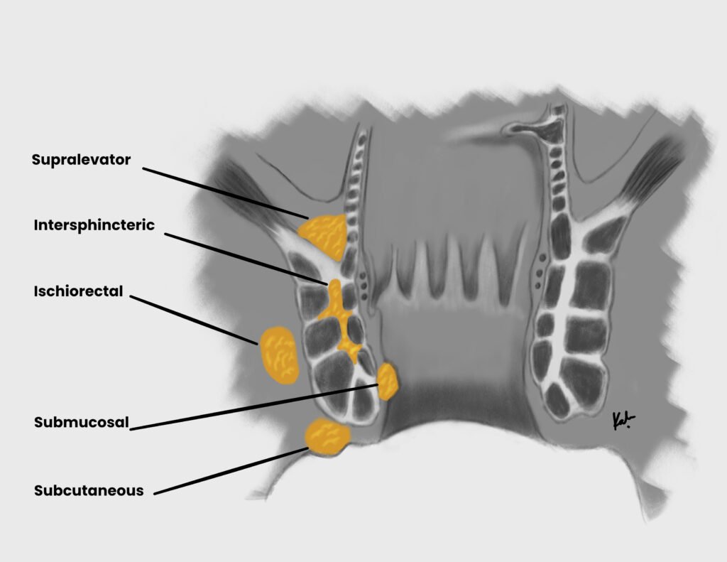
Anatomical locations of anorectal abscess
Anorectal abscess Location Ischiorectal abscess (most common) Abscess beneath the levator ani, traverses the external sphincter into the ischiorectal space. Spreads laterally from the crypts, through the external sphincter and into fatty tissue of ischiorectal space Perianal abscess Abscess beneath the perianal skin , does not traverse the external sphincter. Extends distally from the crypts to skin surrounding anal verge Intersphincteric abscess Abscess between the internal and external sphincter. Confined to crypts and glands. Supralevator abscess (least common) Abscess above the levator ani, extension of intersphincteric abscess OR suppurative abdominal condition (appendicitis, complicated diverticulitis). Extends proximally from crypts past the levator ani muscles into the supralevator space.
Location of anorectal abscesses
Risk factors for anorectal abscess
Flow obstruction and infection of anal crypt glands (MCC, 90%)
Inflammatory bowel disease (chron’s, ulcerative colitis)
Acute appendicitis, Complicated diverticulitis
Radiation-induced proctitis (secondary to radiotherapy for ca prostate)
Iatrogenic (post-hemorrhoidectomy via iatrogenic sinuses or fistulas)
Foreign bodies (ingested toothpick or bones; stuff in anus >.<)
Colorectal cancer
Immunosuppression (may present with leukopenia or perianal pain without noticeable swelling or signs of infection)
Pathophysiology
Obstruction of anal glands by debris → stasis and bacterial overgrowth → Abscess
Extension of abscess into perirectal spaces (intersphincteric, transphincteric, suprasphincteric, extrasphincteric) → fistula formation, bacteremia, sepsis
Signs and symptoms
Constant anorectal pain not associated or worsened with defecation
Swelling at or around the rectum/anus
Fever and chills
Apparent cellulitis or abscess on buttock
Area of focal tenderness on DRE
Erythematous, subcutaneous mass near the anus = perianal abscess
Rectal or perirectal drainage (bloody, purulent, mucoid) = perirectal abscess
Purulent drainage from the anal canal or anoderm = Fistula
DRE findings for anorectal abscess
Fluctuant, indurated mass
Pain with pressure
Investigations
CT or MRI scan: confirm location of deep abscess
Anal ultrasound: confirm deep abscess
Proctosigmoidoscopy or anoscopy: rule out other comorbidities (IBD , Malignancy)
Fistula probe with methylene blue: identify possible fistulae and comorbidities (IBD , Malignancy)
Treatment
Incision and drainage: stops further infection and tissue damage (can lead to sphincter damage, promote fistula formation, and lead to sepsis )
Sitz bath
Analgesics and stool softeners
Post-op antibiotics: ciprofloxaxin and metronidazole to cover gram-negatives and anerobes.
Indications for post-operative antibiotics
Immunocompromised patients
Diabetic patients
Patients with valvular heart disease
Biochemistry
ACTH P: <80 ng/L
ALT P: 5–35 U/L
Albumin P: 35–50 g/L
Aldosterone P: 100–500 pmol/L
Alk. phosphatase P: 30–130 U/L
α-Amylase P: 0–180 IU/dL
α-Fetoprotein S: <10 kU/L
Angiotensin II P: 5–35 pmol/L
ADH P: 0.9–4.6 pmol/L
AST P: 5–35 U/L
Bicarbonate P: 24–30 mmol/L
Bilirubin P: 3–17 μmol/L
BNP P: <50 ng/L
CRP P: <10 mg/L
Calcitonin P: <0.1 mcg/L
Calcium (ionized) P: 1.0–1.25 mmol/L
Calcium (total) P: 2.12–2.60 mmol/L
Chloride P: 95–105 mmol/L
Cholesterol P: <5.0 mmol/L
VLDL P: 0.128–0.645 mmol/L
LDL P: <2.0 mmol/L
HDL P: 0.9–1.93 mmol/L
Cortisol AM P: 450–700 nmol/L
Cortisol Midnight P: 80–280 nmol/L
CK ♂ P: 25–195 U/L
CK ♀ P: 25–170 U/L
Creatinine P: 70–100 μmol/L
Ferritin P: 12–200 mcg/L
Folate S: 2.1 mcg/L
FSH P: 2–8 U/L ♂; >25 menopause
GGT ♂ P: 11–51 U/L
GGT ♀ P: 7–33 U/L
Glucose (fasting) P: 3.5–5.5 mmol/L
Growth hormone P: <20 mu/L
HbA1C (DCCT) B: 4–6%
HbA1C (IFCC) B: 20–42 mmol/mol
Iron ♂ S: 14–31 μmol/L
Iron ♀ S: 11–30 μmol/L
Lactate (venous) P: 0.6–2.4 mmol/L
Lactate (arterial) P: 0.6–1.8 mmol/L
LDH P: 70–250 U/L
LH P: 3–16 U/L
Magnesium P: 0.75–1.05 mmol/L
Osmolality P: 278–305 mosmol/kg
PTH P: 0.8–8.5 pmol/L
Potassium P: 3.5–5.3 mmol/L
Prolactin ♂ P: <450 U/L
Prolactin ♀ P: <600 U/L
PSA P: 0–4 mcg/mL
Protein (total) P: 60–80 g/L
Red cell folate B: 0.36–1.44 μmol/L
Renin (erect) P: 2.8–4.5 pmol/mL/h
Renin (recumbent) P: 1.1–2.7 pmol/mL/h
Sodium P: 135–145 mmol/L
TBG P: 7–17 mg/L
TSH P: 0.5–4.2 mU/L
T4 P: 70–140 nmol/L
Free T4 P: 9–22 pmol/L
TIBC S: 54–75 μmol/L
Triglycerides P: 0.50–2.3 mmol/L
T3 P: 1.2–3.0 nmol/L
Troponin T P: <0.1 mcg/L
Urate ♂ P: 210–480 μmol/L
Urate ♀ P: 150–390 μmol/L
Urea P: 2.5–6.7 mmol/L
Vitamin B12 S: 0.13–0.68 nmol/L
Vitamin D S: 50 nmol/L
Arterial Blood Gases
pH 7.35–7.45
PaCO₂ 4.7–6.0 kPa
PaO₂ >10.6 kPa
Base excess ±2 mmol/L
Urine
Cortisol (free) <280 nmol/24h
Hydroxyindole acetic acid 16–73 μmol/24h
Hydroxymethylmandelic acid 16–48 μmol/24h
Metanephrines 0.03–0.69 μmol/mmol cr.
Osmolality 350–1000 mosmol/kg
17-Oxogenic steroids ♂ 28–30 μmol/24h
17-Oxogenic steroids ♀ 21–66 μmol/24h
17-Oxosteroids ♂ 17–76 μmol/24h
17-Oxosteroids ♀ 14–59 μmol/24h
Phosphate (inorganic) 15–50 mmol/24h
Potassium 14–120 mmol/24h
Protein <150 mg/24h
Protein/creatinine ratio <3 mg/mmol
Sodium 100–250 mmol/24h
Haematology
WCC 4.0–11.0 ×10⁹/L
RBC ♂ 4.5–6.5 ×10¹²/L
RBC ♀ 3.9–5.6 ×10¹²/L
Hb ♂ 130–180 g/L
Hb ♀ 115–160 g/L
PCV ♂ 0.4–0.54 L/L
PCV ♀ 0.37–0.47 L/L
MCV 76–96 fL
MCH 27–32 pg
MCHC 300–360 g/L
RDW 11.6–14.6%
Neutrophils 2.0–7.5 ×10⁹/L (40–75%)
Lymphocytes 1.0–4.5 ×10⁹/L (20–45%)
Eosinophils 0.04–0.44 ×10⁹/L (1–6%)
Basophils 0–0.10 ×10⁹/L (0–1%)
Monocytes 0.2–0.8 ×10⁹/L (2–10%)
Platelets 150–400 ×10⁹/L
Reticulocytes 0.8–2.0% / 25–100 ×10⁹/L
Prothrombin time 10–14 s
APTT 35–45 s
Paediatric
Pulse Rate (bpm)
Neonate 140–160
Infant <1yr 120–140
1–5 years 110–130
5–12 years 80–120
>12 years 70–100
Respiratory Rate (tachypnoea)
0–2 months ≥60/min
2–12 months ≥50/min
1–5 years ≥40/min
>5 years ≥30/min
Blood Pressure (mmHg)
Term 65/45
1 year 75/50
4 years 85/60
8 years 95/65
10 years 100/70
Weight Formulas
3–12 months (a + 9)/2 kg
1–6 years 2a + 8 kg
>6 years (7a − 5)/2 kg
Haemoglobin (g/dL)
Term newborn 13–20
1 month 11–18
2 months 10–15
1–2 years 10–13
>2 years 11–14
MUAC (6 months–5 years)
Obese >17.5 cm
Normal 13.5–17.4 cm
At risk 12.5–13.4 cm
Moderate malnutrition 11.5–12.4 cm
Severe malnutrition <11.5 cm
Developmental Milestones
Social smile 1.5 months
Head control 4 months
Sits unsupported 7 months
Crawls 10 months
Stands unsupported 10–12 months
Walks 12–13 months
Talks 18 months
CSF WBC (/mm³)
Term newborn 0–25
>2 weeks 0–5