A 2-day-old female infant born at 39 weeks via breech vaginal delivery is undergoing her routine newborn exam on day 2 of life. The baby is feeding well, active and has no apparent distress.
On examination, she is a healthy term neonate. There is a positive Barlow test and a positive Ortolani test on the left. There is no leg length discrepancy. No other congenital anomalies are noted.
Q1. What is the diagnosis?
Reveal answer
Developmental dysplasia of the hip (DDH)
DDH is an abnormal development of the hip characterised by dysplasia, subluxation and possible dislocation of the hip. It is the most common orthopaedic disorder in newborns.
Q2. What are the risk factors for this condition?
Reveal answer
Firstborn child (due to tight spacing in the uterine cavity)
Female (due to ligamentous laxity caused by circulating estrogens)
Family history of DDH (parents or siblings)
Fluid lacking (oligohydramnios)
Feet first (breech presentation especially when feet lie near the shoulders e.g. Frank breech)
Tight swaddling (especially with the hips and knees straight)
5 F’s: firstborn, female, family history, fluid lacking, feet first
Q3. What associated conditions should be looked for?
Reveal answer
Congenital muscular torticollis
Metatarsus adductus
Congenital knee dislocation
These condition can also be caused by intrauterine malpositioning – packaging disorders
Q4. What is the pathophysiology of this condition and how does it relate to its risk factors?
Reveal answer
Maternal and fetal ligamentous laxity due to hormonal influence (estrogen and relaxing) leads to intrauterine malpositioning
Mechanical factors (breech presentation, oligohydramnios, primigravida with reduced intrauterine space) also leads to intra-uterine malpositioning
Post-natal positioning (sweight swaddling with hip extended and adducted) exacerbates this instability
Due to this malpositioning there is poor contact between the femoral head and acetabulum leading to deficiency of the anterior and anterolateral acetabulum (the socket is shallow and unstable)
Bony changes:
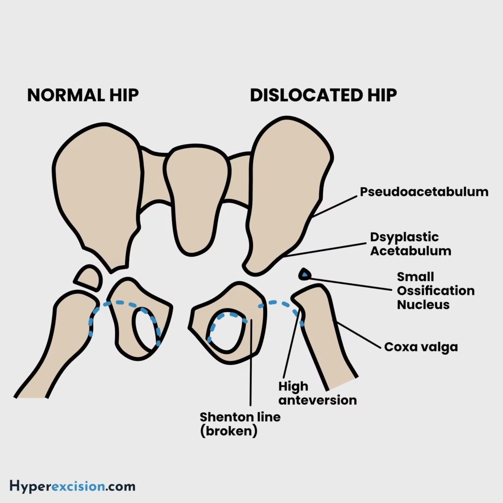
Shallow and dysplastic acetabulum
Anteverted and anterolaterally deficient acetabulum
Pseudoacetabulum formation: the femoral head may articulate with the ilium at a false socket
Delay in teardrop development
Delayed ossific nucleus of the femur, anteversion, deformed head and coxa valgus
Intra-articular soft tissue changes:
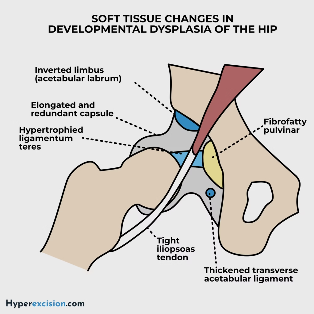
Elongated and redundant capsule.
Thickened ligamentum teres
Thickened transverse acetabular ligament
Limbus formation as the displaced femoral head causes hypertrophy and inversion of the acetabular cartilage rim (creates a physical barrier to reduction)
Pulvinar muscles
Extra-articular soft tissue changes:
Tight adductors
Tight psoas tendon
Short abductors
Q5. What are the phases (classification) of this condition?
Reveal answer
| Phase | Physical exam |
|---|---|
| Subluxable | Barlow-suggestive |
| Dislocatable | Barlow-positive |
| Dislocated | Ortolani-positive if reducible, Ortolani negative if irreducible |
Q6. What signs can be elicited during physical examination in this condition?
Reveal answer
| Age | Signs |
|---|---|
| < 3 months | Positive barlow, positive ortolani, Galeazzi (allis test), telescoping, asymmetric skin folds |
| 3 – 12 months | Decreased hip abduction (due to contracture), leg length discrepancy, Klisic test (for bilatral dislocation) |
| > 12 months (walking child) | Pelvic obliquity, lumbar lordosis (due to hip contractures), trendelenburg gait (due to abductor insufficiency), toe-walking (to compensate for shortening of the affected side) |
Q7. Describe the following tests: Barlow, Ortolani, Galeazzi (Allis), Klisic
Reveal answer
| Test | Purpose | Maneuver | Positive finding |
|---|---|---|---|
| Barlow | Dislocatable hip | Adduction + posterior pressure | Hip dislocates (”clunk”) |
| Ortolani | Reducible hip | Abduct + anterior lift | Hip reduces (”clunk”) |
| Galeazzi (Allis) | Leg length discrepancy | Inspect with knees flexed at 90 degrees | One knee is lower |
| Klisic | Chronic dislocation | Line drawn from the ASIS to trochanter line | Points below the umbilicus |
Q8. Which radiographic reference lines are used to assess normal hip alignment on a paediatric pelvic radiograph?
Reveal answer
| Line | Description | Importance / Use |
|---|---|---|
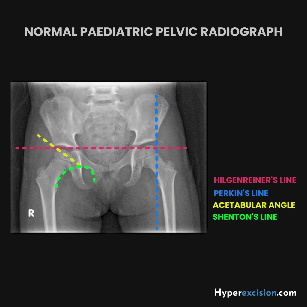
| Hilgenreiner’s line | Horizontal line through triradiate cartilages (Y-shaped growth plate) | Baseline for measuring acetabular angle; used in developmental dysplasia of the hip (DDH) |
| Perkins’ line | Vertical line at lateral edge of acetabulum, perpendicular to Hilgenreiner’s | Defines quadrants; femoral head should lie in inferomedial quadrant (displacement = DDH) |
| Shenton’s line | Smooth arc along inferior border of superior pubic ramus → medial femoral neck | Interruption suggests hip dislocation, subluxation, or fracture |
| Acetabular angle | Angle between Hilgenreiner’s line & acetabular roof line | Increased angle indicates acetabular dysplasia |
| Klein’s line | Line along superior border of femoral neck; should intersect femoral epiphysis | Failure = slipped capital femoral epiphysis (SCFE) |
Q9. What investigations are useful for this condition?
Reveal answer
Ultrasound of the hip: for patients less than 6 months old
Acetabular dysplasia
Hip dislocation
X-ray of the hip: AP view of pelvis for patients older than 6 months when the ossific nucleus of the proximal femur appears
Shenton’s line (should be continuous)
Hilgenreiner’s line (femoral head ossification should be inferior to this line)
Perkin’s line (femoral head ossification should be medial to this line
Delayed ossification of the femoral head
Absent acetabular teardrop
Arthrogram: to confirm reduction after closed reduction under anaesthesia
MRI: to evaluate reduction of hip after closed reduction and spica casting
Q10. How is this condition treated?
Reveal answer
| Treatment | Description | Nota bene |
|---|---|---|
| Pavlik harness | For children less than 6 months old | Assess every 4 – 6 weeks with ultrasound. Assess after 2 weeks if reduced → If reduced wear for 6 weeks → confirm after 6 weeks if reduced → if reduced wean off over 6 – 8 weeks. |
| Closed manipulative reduction (CMR) and hip spica | For children 6 – 18 months old | Reduction is confirmed using an arthrogram. Spica is changed every 6 weeks and worn for a total of 3 months. |
| Operative treatment | For children older than 18 months | Includes open reduction +/- femoral/pelvic osteotomy |
Q11. What are the principles of using a Pavlik harness?
Reveal answer
Dynamic splint (requires normal muscle function – works as the child kicks)
Use before the child can sit
Anterior strap flexes the hip to 90 – 100 degrees while the posterior strap abducts the hip to 45 degrees
Worn for 24 hours a day for 6 weeks
Q12. What advice can be given to parents regarding the hip spica?
Reveal answer
Small and frequent meals: to prevent bloating and constipation which can make the child uncomfortable in the cast
Perineal care: clean and dry perineum to prevent rashes and infection
Cast care: the cast should be dry, clean and intact all times. Use sponge bath only.
Q13. What is the safe zone of Ramsey?
Reveal answer
The safe zone of ramsey is the zone between maximum abduction and minimum adduction where the hip remains reduced
It should be at least 25 degrees. The wider the zone of safety the more stable the reduction.
If the safe zone of ramsey in children being treated for DDH with a spica is not more than 25 degrees adductor tenotomy can be performed
Q14. What complications may occur if this condition is untreated or poorly treated?
Reveal answer
Pavlik harness disease: erosion of the posterior acetabulum due to prolonged use of the Pavlik harness with the hip dislocated
Femoral nerve palsy: due to overflexion when using a Pavlik harness
Avascular necrosis of the femoral head: due to over-abduction > 60 degrees when using a Pavlik harness. This causes impingement of the retinacular branch of the medial femoral circumflex artery.
Recurrence (10%)
Chronic limp
Limb length discrepancy which leads to back pain, functional scoliosis and knee pain
Pain or decreased range of motion