A 14-year-old boy presents with a 3-month history of progressive pain and swelling around his left knee. The pain is dull, constant, and often wakes him at night. His parents report that he has developed a limp and is unable to participate in sports. He has also experienced some weight loss and fatigue, though there has been no history of fever or night sweats.
Q1. What warning signs suggest that this child might have a bone tumour?
Unremitting bone or joint pain (especially pain that wakes the patient from sleep)
Rapid progression of bone or joint pain/swelling
Constitutional symptoms such as weight loss
Q2. What is the differential diagnosis for a bone tumour in children?
Osteogenic sarcoma
Ewing’s sarcoma
Osteogenic sarcoma is the most common primary bone tumour in individuals < 30 years. It is seen in older children and young adults aged 10 – 25 years (75%), with a second peak in older adults aged 60 – 79 years (secondary to radiation or Paget’s disease)
Q3. What are the most common bone tumours by age group?
Most common bone tumors by age group
| Age group | Common tumors |
|---|---|
| Paediatric | Ewing sarcoma, osteogenic sarcoma (osteosarcoma) |
| Adult | Metastasis to bone, multiple myeloma, lymphoma, sarcoma of soft tissue |
Q4. List the most common bone tumours by location?
Bone tumours by location
| Location | Tumors |
|---|---|
| Skull | Eosinophilic granuloma, metastasis, myeloma |
| Mandible | Adamantinoma, dentogenic cyst |
| Humerus | All tumors, simple bone cysts |
| Ribs | Metastases, enchondroma, chondrosarcoma, Ewing’s sarcoma |
| Spine | Metastases, myeloma, osteoblastoma, chordoma, hemangioma, aneurysmal bone cyst |
| Pelvis | Osteosarcoma, chondrosarcoma, simple bone cyst, lymphoma, Ewing’s sarcoma, AND chordoma |
| Distal radius | Giant cell tumor, eosinophilic granuloma |
| Hands | Enchondroma |
| Proximal femur | Simple bone cyst, fibrous dysplasia, enchondroma, Ewing sarcoma, eosinophilic granuloma |
| Around the knee | All tumors, osteosarcoma, Ewing’s sarcoma |
| Tibia shaft | Non-ossifying fibroma, fibrous dysplasia, adamantinoma, osteoid osteoma |
| Calcaneus | Simple bone cyst, Ewing’s sarcoma, osteoblastoma, chondroblastoma |
On physical examination, there is a firm, tender swelling over the distal femur measuring approximately 6 cm. The swelling is immobile and associated with warmth and restriction of knee movement. The overlying skin appears stretched but intact, with no redness or ulceration. There is no palpable lymphadenopathy, and distal pulses are well felt.
Laboratory studies show the following results:
| Test | Result | Reference Range | Interpretation |
|---|---|---|---|
| Hemoglobin | 10.5 g/dL | 12–16 g/dL | Mild anemia |
| ESR | 40 mm/hr | <20 mm/hr | Elevated |
| CRP | Normal | <10 mg/L | Normal |
| Alkaline Phosphatase (ALP) | 700 U/L | <150 U/L | Markedly elevated |
| Lactate Dehydrogenase (LDH) | 550 U/L | <250 U/L | Elevated |
A plain X-ray of the distal femur shows a destructive lesion in the metaphyseal region with mixed lytic and sclerotic changes. There is periosteal elevation forming a Codman’s triangle, and a characteristic “sunburst” appearance of periosteal reaction. Cortical breach with extension into adjacent soft tissue is also seen. MRI confirms a large heterogeneous metaphyseal mass extending into surrounding soft tissue, but without encasement of the neurovascular bundle. A staging CT of the chest reveals small pulmonary nodules suggestive of metastases.
A core needle biopsy demonstrates malignant spindle-shaped cells producing osteoid, confirming the diagnosis of osteogenic sarcoma (osteosarcoma) of the distal femur with pulmonary metastases
Q5. What risk factors are associated with osteosarcoma?
Paget’s disease
Radiation
Bone infarction
Li-fraumeni syndrome
Retinoblastoma
Q6. Regarding osteogenic sarcoma, what investigations are useful to make a diagnosis and what is the clinical utility or the expected findings in each investigation?
X-ray of the affected area: best initial step. Also used for staging
“Sunburst” pattern (looks like cotton balls due to periosteal reaction)
Mass often extends beyond the periosteal margins
Codman triangle (newborn formation in response to periosteal lifting)
MRI: for staging. Delineates the medullary and extra-osseous extension of the tumor
CT-scan of chest: rule out lung metastases
Bone scan: evaluate for bone metastases
Biopsy: definitive histological diagnosis
Anaplastic or spindle-shaped cells with nuclear pleomorphism
Islands of osteoid (bone)
Alkaline phosphatase: elevated
Lactate dehydrogenase: elevated
Erythrocyte sedimentation rate: elevated
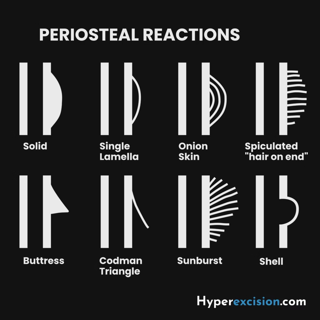
Q7. The characteristic periosteal reaction in osteosarcoma is a “sunburst” pattern or “Codman’s triangle”. What other periosteal reactions can occur with bone tumours?
| Periosteal Reaction | Appearance | Suggestive of |
|---|---|---|
| Solid/Continuous | Single dense layer | Benign, slow-growing (healing fracture, osteoid osteoma) |
| Lamellated (Onion-skin) | Multiple concentric layers | Ewing’s sarcoma, osteomyelitis, lymphoma |
| Spiculated (Hair-on-end / Sunburst) | Radiating spicules | Osteosarcoma, aggressive tumors |
| Codman’s Triangle | Triangular elevation at tumor margin | Aggressive tumors (osteosarcoma, Ewing’s, metastases) |
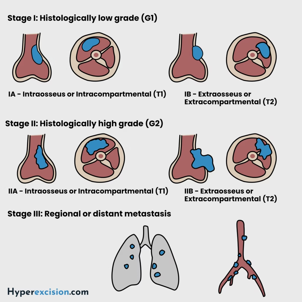
Q8. The MSTS (Enneking) tumour staging system is a widely used framework for staging musculoskeletal tumours (bone and soft tissue sarcomas). What does it entail and how does this relate to treatment?
Enneking staging is based on 3 key factors:
Tumor grade (G): low-grade vs high-grade
Site or local spread (T): intra-compartmental vs extra-compartmental
Metastasis (M): no mets vs distant mets
MSTS (Enneking) staging
| Stage | Grade | Site | Metastasis |
|---|---|---|---|
| IA | Low grade | T1 – intracompartmental | M0 |
| IB | Low grade | T2 – extracompartmental | M0 |
| IIA | High grade | T1 – intracompartmental | M0 |
| IIB | High grade | T2 – extracompartmental | M0 |
| III | Metastatic | T1 or T2 intracompartmental or extracompartmental | M1 |
Summary of staging and treatment
| Stage | Description | Treatment |
|---|---|---|
| Stage I (low-grade) | Grows slowly, rarely metastasizes | Wide local excision (with preservation of limb) |
| Stage II (high-grade) | Grows rapidly with a higher risk of recurrence and metastasis | Wide local excision or radical excision. Limb salvage if clear margins are achievable vs amputation if limb cannot be salvaged. Adjuvant chemotherpay +/- radiotherapy |
| Stage III (metastatic disease) | Poor prognosis. | Multimodal treatment: systemic chemotherapy, surgical resection of primary tumor, possible resection of lung metastases, palliative care |
A compartment is an anatomical space bounded by natural barriers, e.g., bone cortex or fascial envelope. This usually limits the spread of a tumour. For instance, the thigh has an anterior compartment, medial compartment, and posterior compartment based on fascial planes. In addition, each bone is considered a single compartment. For example, if a tumour is confined to the femur without soft-tissue extension, it is still intracompartmental.
Radiotherapy has a limited role in osteosarcoma since it is relatively radioresistant
Q9. What are the principles of surgery for bone tumours?
| Type of Excision | Margin Relative to Tumor | Typical Use |
|---|---|---|
| Intralesional excision | Inside tumor (incomplete) | Benign tumors, palliative care |
| Marginal excision | At the tumor edge / pseudocapsule | Some benign aggressive tumors |
| Wide local excision | Through normal tissue outside the reactive zone (with clear margins, within the same compartment) | Limb-salvage for intra-compartmental tumours or low-grade malignant tumours |
| Radical excision | The entire compartment was removed | For high-grade tumors, recurrence, or when a wide excision is not possible |