A 12-year-old boy was brought to the clinic with 1 1-month history of left groin pain and a limp.
Q1. What is the difference between pain that radiates into the groin and pain that originates in the groin?
Reveal Answer
Pain radiating to the groin suggests urologic, reproductive or spinal etiology
Pain originating in the groin suggests pathology in or around the hip joint
True groin pain is proximal and medial. Clarify whether the hip pain is proximal and lateral vs medial. Pain that is lateral may be due to trochanteric bursitis, neurogenic claudication, sciatica or sacroiliac joint pathology
Similarly, in the shoulder joint, pain from glenohumeral arthritis is felt medially in the axilla, while that from rotator cuff or subacromial bursitis is felt laterally over the shoulder.
Q2. What are the differentials for groin pain with refusal to bear weight in a child?
Reveal Answer
Mechanical
| Differentials | Clinical features |
|---|---|
| Slipped upper femoral epiphysis (SUFE) | Obese adolescent male (10 – 16 years due to rapid growth) with groin pain radiating to the knee, painful limp, and externally rotated hip. May allow range of motion. Affects the left hip more than the right. |
| Perthes disease | Gradual progressive limp with slow onset of pain, decreased range of motion. Peak incidence at 4 – 8 years. |
| Developmental dysplasia of the hip | Positive Barlow and Ortolani, Galeazzi sign, positive Trendelenburg sign, delayed walking or abnormal gait if left untreated. Screening is done at birth and is unusual to be picked up at 12 years old |
| Stress frActure | Pain associated with activity or repeated loading, no history of recent truma, local tenderness and swelling |
Infectious
| Differentials | Description |
|---|---|
| Septic arthritis or osteomyelitis | Fever, pain and inability to move or bear weight, may have underlying joint disease e.g. Rheumatoid arthritis |
| Psoas abscess | Fever, hip flexed and internally rotated, pain on passive hip extension, associated with osteomyelitis of the spine or pelvic inflammatory disease |
Inflammatory
| Differentials | Description |
|---|---|
| Juvenile idiopathic arthritis | Non-migratory arthritis affecting one or more joints for > 3 months, morning stiffness, fever |
| Transient synovitis | Most common cause of hip pain. History of viral infection, hip pain worse on waking and improves as day progresses, self-limiting. Peak incidence at 2 – 5 years (maximum around 12 years). |
Benign neoplasm
| Differentials | Description |
|---|---|
| Osteoid osteoma | Continous, deep, aching bone pain affecting the neck or back; unexplained, rigid, painful scoliosis |
Malignant neoplasm
| Differential | Description |
|---|---|
| Osteosarcoma | Most common primary malignant bone tumour. Common in adolescents. Presents with deep bone pain that progresses to palpable bony mass. Affects the distal femur |
| Ewing sarcoma | Affects children aged 5 – 15 years, bone pain, fever, fatigue, weight loss, pathologic fractures and a palpable mass |
The most important differential is septic arthritis since it can cause irreversible damage to cartilage. History of fever, refusal to bear weight and laboratory values (+ Kocher criteria) can be used to confirm this diagnosis.
Q3. What questions are important to ask for in this child’s history?
Reveal Answer
History of fever (to rule out infection e.g. septic arthritis)
Recent history of viral illness (to rule out transietn synovitis)
Family history of joint disease (to rule out conditions such ad DDH and juvenile idiopathic arthritis)
Medications (a history of steroid use can predispose to perthes disease)
The pain began in his left groin and radiated to the left knee. It has been gradually increasing in intensity. It is worse with walking and relieved by rest. There is no history of trauma. He does not have pain in other joints or extremities. There is no history of recent infection, no history of fever, chills or malaise. He participates in physical education in school but is otherwise not involved in sports. He has no history of travel or camping trips and lives in an urban area. He takes no medication. There is no family history of joint problems.
On physical examination, he is afebrile and appears obese. The left lower extremity is externally rotated, abducted and flexed, and he resists internal rotation. There is no leg length discrepancy. Neurologic and vascular examination is normal.
Q4. What are the steps in the orthopaedic examination of a painful hip?
Reveal Answer
Look (inspection – attitude and leg length)
Feel (palpation of the ASIS, trochanteric bursa and muscle insertions)
Move (active and passive range of motion)
Neurologic (motor and sensory)
Vascular (pulses)
Special tests
Q5. List three special tests used to examine the hip
Reveal Answer
| Test | Description |
|---|---|
| Thomas test | Test for flexion contracture. With the patient lying suine one knee is brougth to the chest. If the opposite leg is unable to remain extended there is flexion contracture of the hip |
| Trendelenburg test | Patient stands in position and lifts one leg. If the pelvis falls on the side of the raised leg there is abductor weakness on the side of the standing leg (contralateral) |
| FABER | Flexion, abduction and external rotation of the hip with the patient lying supine. Pain during this movement suggests sacroiliac joint pathology. |
Q6. What is the most likely diagnosis?
Reveal Answer
Slipped upper femoral epiphysis (SUFE) affecting the right lower extremity
Age 12 years (period of rapid growth)
Non-traumatic groin pain radiating to the knee
External rotation and abduction of the hip with flexion (Drehmann’s sign)
Resistance to internal rotation
SUFE is displacement of the metaphysis of the femoral neck from teh epiphysis through the growth plate (physis). Commonly due to overloading of teh physis (oesity) or weakened growth plate (endocrine disorder). The neck displaces anteriorly and superiorly relative to teh femoral epiphysis
External rotation of the leg is the most common resting position in SUFE because of the mechanical direction of slippage. Obligate external rotation can be observed when the hip is flexed, i.e. the leg involuntarily externally rotates
Q7. External rotation of the leg is the most common resting position in SUFE. What should be considered if the limb is held in slight hip flexion and internally rotated instead?
Reveal Answer
Psoas abscess
Hip dislocation Psoas abscess is associated with fever and other signs of infection. The source of the abscess should be investigated e.g. osteomyelitis of the spine, pelvic inflammAtory disease An internally rotated and flexed hip following trauma is hip dislocation until proven otherwise. This is a surgical emergency.
Q8. In the setting of knee pain, why should the hip be examined?
Reveal Answer
Hip pathology (SUFE and perthes disease) can present with referred pain to the knee
In children, a complaint of knee pain should lead to clinical examination of the hip and a radiograph of the hip
Q9. What are the risk factors for developing SUFE?
Reveal Answer
Age 10 – 16 years
Male gender
Obesity (one of the strongest risk factor due to increased load across the physis increasing the likelihood of slippage)
50% of patients with SUFE are ≥ 90th percentile for weight.
Q10. What condition may be associated with SUFE?
Reveal Answer
Renal osteodystrophy
Excessive growth hormone
Hyper- or hypoparathyroidsm
These conditions physiologically weaken the physis, leading to slippage
With these conditions, SUFE can present early (< 10 years) or later (> 16 years) in life
Suspect endocrine disease in young thin patients (delayed growth or puberty)
Q11. What investigations are useful in this case?
Reveal Answer
Pelvic X-ray (AP and frog-leg lateral view) +/- knee radiographs
Complete blood count and CRP: to rule out infection (septi carthritis)
Blood culture and joint aspiration: if there is suspiscion of infection
Knee radiographs (AP, lateral and sunrise view) can be added if knee pathology is suspected
MRI can be used to rule out non-displaced stress fractures, early necrosis in Perthes disease, soft tissue abnormalities of the ligaments, tendons and labrum, and bone tumours.
CT is used sparingly due to the risk of unnecessary exposure to radiation
A bone scan can be used for osteomyelitis or a tumour
Q12. What are the classic radiographic findings in SUFE vs DDH vs Perthes disease?
Reveal Answer
| Condition | Radiographic findings |
|---|---|
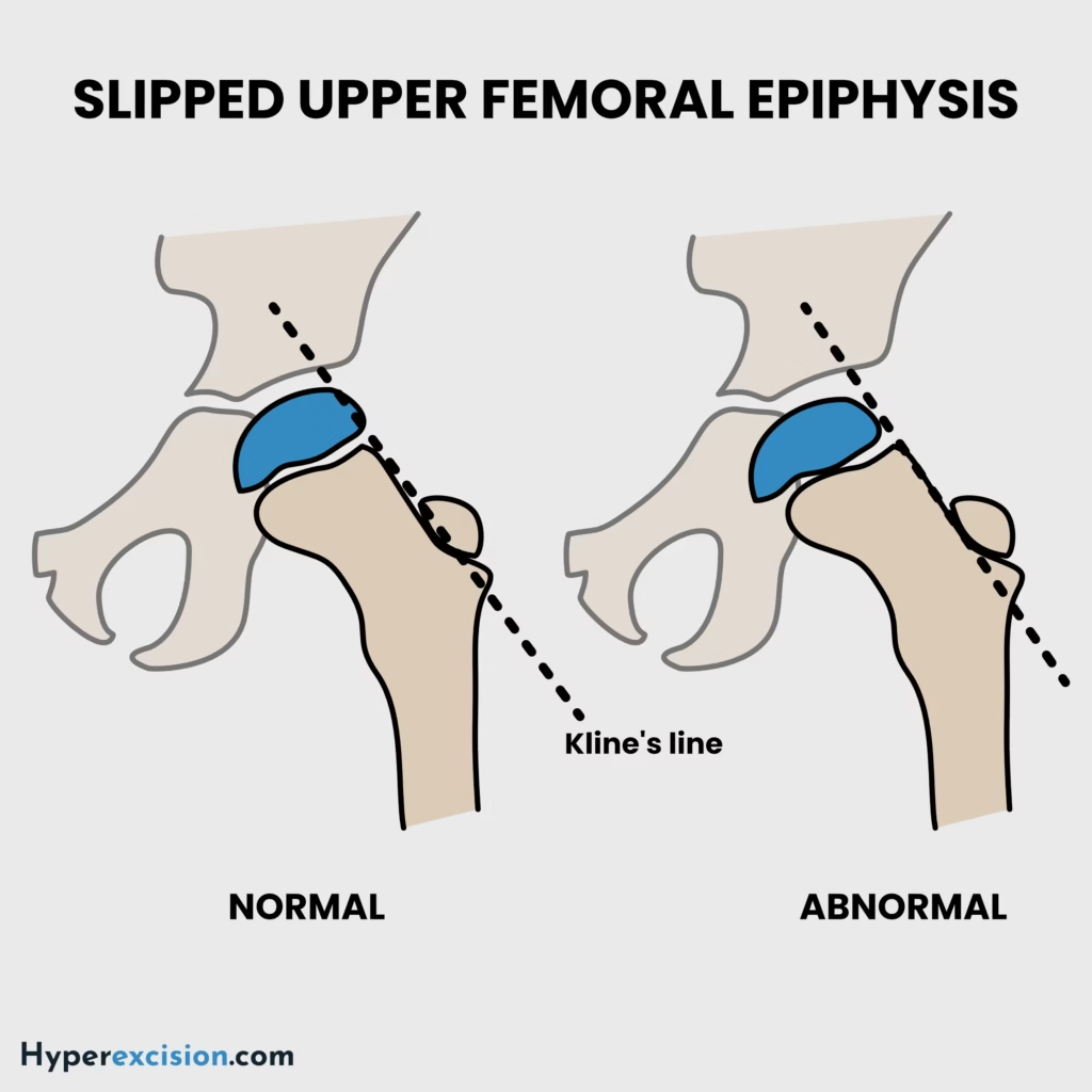
| Slipped upper femoral epiphysis (SUFE) | Klein’s line does not intersect the femoral head, growth plate widening or lucency (epiphysiolysis), blurring of the femoral metaphysis (metaphyseal blanch sign of steel) |
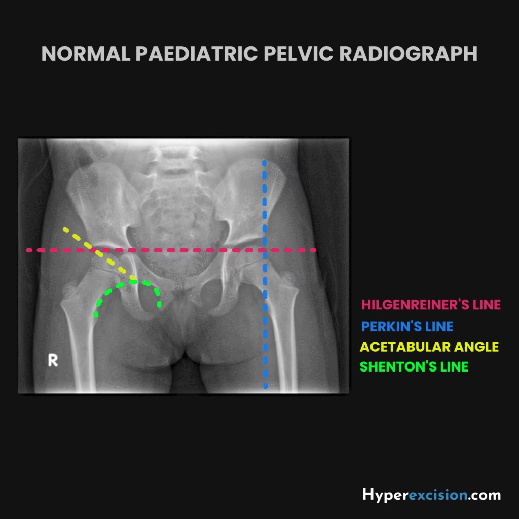
| Developmental dysplasia of the hip (DDH) | Difficult to assess on X-ray for infants but – disrupted shenton’s line, femoral head ossification superior to Hilgenreiner’s line, femoral head ossification lateral to perkin’s line, delayed ossification of teh femoral head, absent acetabular teardrop |
| Perthes disease | Subchondral collapse, flattening and fragmentation of the femoral head |
Q13. What is the treatment of SUFE vs DDH vs Perthes disease?
Reveal Answer
| Condition | Treatment |
|---|---|
| Slipped upper femoral epiphysis (SUFE) | Percutaneous in situ screw fixation through the femoral neck, contralateral in sity prophylactic pinning (bilateral in situ fixation) |
| Developmental dysplasia of the hip (DDH) | Abduction bracing with a Pavlik harnss for < 6 months, open/closed recution and spica cast for 6 – 18 mothns, open reduction and pelvic osteotomy for > 18 months |
| Perthes disease | Most require supportive treatment, advanced disease can be contained with abduction bracing or pelvic/femoral osteotomies |
Q14. What are the complications of SUFE?
Reveal Answer
Osteonecrosis of the femoral head
Contralateral SCFE/SUFE
Chondrolysis
Slip progression
Infection
Chronic pain
Degenerative arthritis
Pin associated proximal femur fracture
Labral tearing and degeneration