Ventilator-Associated Lung Injury (VALI)

Knowledge Check

Exam-style MCQs aligned with these notes. Available to Hyperexcision Scholar members.

Log in to access
Last updated: September 28, 2026Bookmark

Overview

Ventilator-associated Lung Injury (VALI) is acute lung injury caused or worsened by mechanical ventilation. ****It can be caused by both invasive and non-invasive mechanical ventilation.

  • Signs and symptoms
    • Worsening hypoxia
    • Falling SpO2 or PaO2
    • New bilateral infiltrates
    • Evidence of alveolar overdistension or atelectasis
    • Deterioration despite ventilation

Mechanisms of VALI

MechanismDescription
Oxygen toxicityReactive oxygen species (ROS) are produces during prolonged exposure to high concentrations of oxygen. An FiO2 of < 60% should be targeted.
Microbarotrauma (Volutrauma)High tidal volumes excessively stretches alveoli, even in the setting of normal airway pressures.
MacrobarotraumaPressure-related overdistension which leads to pneumothorax, pneumomediastium, and subcutaneous emphysema. A key lung protective strategy is Pplat < 28 – 30 cmH20 in ARDS
Repeated alveolar collapse and expansion (RACE or Atelectotrauma)Repeated alveolar opening and closing causes shear stress. An appropriate PEEP helps to keep alveoli open, but excessive PEEP can cause overdistension.
BiotraumaMechanical injury causes inflammatory mediators to be released leading to systemic inflammation and multiorgan failure
Effort-induced injuryExcessive spontaneous respiratory effort generates injurious transpulmonary pressure
Deflation InjuryAbrupt loss of PEEP can cause atelectasis and pulmonary oedema

Driving Pressure

Driving pressure is the pressure that is applied to the alveoli to expand them during inspiration.

A higher driving pressure is associated with mortality in ARDS and is an important marker of ventilator-induced stress.

Physiologically, driving pressure is the ratio of tidal volume (CT) to respiratory system compliance (Crs)
Driving pressure can be easily calculated at the bedside using Pplat – PEEP in a patient that is passive on the ventilator. Alveolar pressure rises from baseline (PEEP) at the end of expiration up to a maximum static stretch (Pplat) at the end of inspiration

Parameter and targets

ParameterTarget
Driving pressure (delta P)< 15 cmH20. A higher driving pressure is associated with VALI.
Pplat< 30 cmH20. A high Pplat is associated with barotrauma and overdistension.
  • Driving pressure and ARDS
    • The aerated functional parts of the lungs in ARDS is greatly reduced. This is known as the ‘baby lung’ concept.
    • The ‘baby lung’ is small with reduced compliance. Pushing the same 6 – 8 ml/kg tidal volume into a lung with low-compliance causes a high driving pressure and greatly overdistends the lungs.
    • A higher PEEP in ARDS recruits collapsed alveoli, improves lung compliance and reduces driving pressure.
    • A lower tidal volume in ARDS also decrease driving pressure, with some permissive hypercapnia (provided the pH of the patient remains acceptable).

Pressure-Volume (PV) Curve

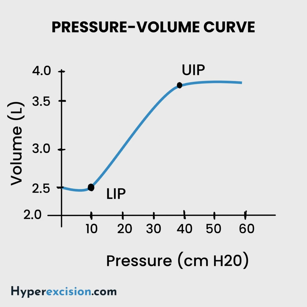
The pressure-volume curve of the respiratory system is sigmoidal with a lower inflexion point (LIP) and upper inflexion point. The ventilation range should be between the LIP and UIP

Below LIP – alveoli collapse → repeated opening/closing → RACE/atelectotrauma

Above UIP – alveoli are overdistended → microbarotrauma and macrobarotrauma

Pressure volume curve
  • PEEP and the PV curve
    • PEEP determines the end-expiratory pressure and volume
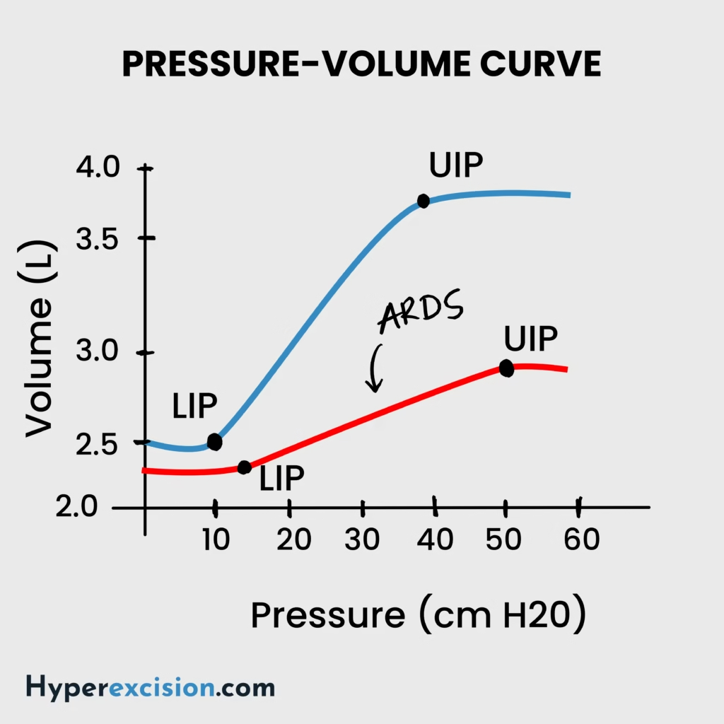
  • ARDS and the PV curve
    • ARDS causes the PV curve to shift downwards and to the right since lung compliance is reduced
    • Thus the same ventilator setting produces higher plateau pressures after a patient develops ARDS
The pressure volume curve in ARDS

Prevention of VALI

VALI can be prevented by avoiding alveolar overdistension and repeated alveolar collapse.

  1. Low tidal volume strategy
    1. VT ~ 4-6 ml/kg predicted body weight in ARDS
    2. VT ~ 6-8 ml/kg predicted body weight in non-ARDS
  2. Control Plateau pressure
    1. Pplat < 30 cmH20 to reduce alveolar overdistension and barotrauma
  3. Optimize PEEP
    1. Too little PEEP = alveolar collapse → RACE
    2. Too much PEEP = overdistension and reduced preload/increased afterload → barotrauma and haemodynamic compromise
    3. Use closed suction systems in ARDS since open suction can cause a sudden loss of PEEP and derecruitement
  4. Prone positioning
    1. Proning in ARDS with PaO2/FiO2 < 150 (PROSEVA trial)
  5. Neuromuscular blockade
    1. This reduces excessive patient effort and effort-induced injury (ACURASYS study)
  6. Avoid excessive FiO2
    1. Aim to titrate FiO2 to < 60% as soon as possible

Reference Intervals ›
Biochemistry
ACTHP: <80 ng/L
ALTP: 5–35 U/L
AlbuminP: 35–50 g/L
AldosteroneP: 100–500 pmol/L
Alk. phosphataseP: 30–130 U/L
α-AmylaseP: 0–180 IU/dL
α-FetoproteinS: <10 kU/L
Angiotensin IIP: 5–35 pmol/L
ADHP: 0.9–4.6 pmol/L
ASTP: 5–35 U/L
BicarbonateP: 24–30 mmol/L
BilirubinP: 3–17 μmol/L
BNPP: <50 ng/L
CRPP: <10 mg/L
CalcitoninP: <0.1 mcg/L
Calcium (ionized)P: 1.0–1.25 mmol/L
Calcium (total)P: 2.12–2.60 mmol/L
ChlorideP: 95–105 mmol/L
CholesterolP: <5.0 mmol/L
VLDLP: 0.128–0.645 mmol/L
LDLP: <2.0 mmol/L
HDLP: 0.9–1.93 mmol/L
Cortisol AMP: 450–700 nmol/L
Cortisol MidnightP: 80–280 nmol/L
CK ♂P: 25–195 U/L
CK ♀P: 25–170 U/L
CreatinineP: 70–100 μmol/L
FerritinP: 12–200 mcg/L
FolateS: 2.1 mcg/L
FSHP: 2–8 U/L ♂; >25 menopause
GGT ♂P: 11–51 U/L
GGT ♀P: 7–33 U/L
Glucose (fasting)P: 3.5–5.5 mmol/L
Growth hormoneP: <20 mu/L
HbA1C (DCCT)B: 4–6%
HbA1C (IFCC)B: 20–42 mmol/mol
Iron ♂S: 14–31 μmol/L
Iron ♀S: 11–30 μmol/L
Lactate (venous)P: 0.6–2.4 mmol/L
Lactate (arterial)P: 0.6–1.8 mmol/L
LDHP: 70–250 U/L
LHP: 3–16 U/L
MagnesiumP: 0.75–1.05 mmol/L
OsmolalityP: 278–305 mosmol/kg
PTHP: 0.8–8.5 pmol/L
PotassiumP: 3.5–5.3 mmol/L
Prolactin ♂P: <450 U/L
Prolactin ♀P: <600 U/L
PSAP: 0–4 mcg/mL
Protein (total)P: 60–80 g/L
Red cell folateB: 0.36–1.44 μmol/L
Renin (erect)P: 2.8–4.5 pmol/mL/h
Renin (recumbent)P: 1.1–2.7 pmol/mL/h
SodiumP: 135–145 mmol/L
TBGP: 7–17 mg/L
TSHP: 0.5–4.2 mU/L
T4P: 70–140 nmol/L
Free T4P: 9–22 pmol/L
TIBCS: 54–75 μmol/L
TriglyceridesP: 0.50–2.3 mmol/L
T3P: 1.2–3.0 nmol/L
Troponin TP: <0.1 mcg/L
Urate ♂P: 210–480 μmol/L
Urate ♀P: 150–390 μmol/L
UreaP: 2.5–6.7 mmol/L
Vitamin B12S: 0.13–0.68 nmol/L
Vitamin DS: 50 nmol/L
Arterial Blood Gases
pH7.35–7.45
PaCO₂4.7–6.0 kPa
PaO₂>10.6 kPa
Base excess±2 mmol/L
Urine
Cortisol (free)<280 nmol/24h
Hydroxyindole acetic acid16–73 μmol/24h
Hydroxymethylmandelic acid16–48 μmol/24h
Metanephrines0.03–0.69 μmol/mmol cr.
Osmolality350–1000 mosmol/kg
17-Oxogenic steroids ♂28–30 μmol/24h
17-Oxogenic steroids ♀21–66 μmol/24h
17-Oxosteroids ♂17–76 μmol/24h
17-Oxosteroids ♀14–59 μmol/24h
Phosphate (inorganic)15–50 mmol/24h
Potassium14–120 mmol/24h
Protein<150 mg/24h
Protein/creatinine ratio<3 mg/mmol
Sodium100–250 mmol/24h
Haematology
WCC4.0–11.0 ×10⁹/L
RBC ♂4.5–6.5 ×10¹²/L
RBC ♀3.9–5.6 ×10¹²/L
Hb ♂130–180 g/L
Hb ♀115–160 g/L
PCV ♂0.4–0.54 L/L
PCV ♀0.37–0.47 L/L
MCV76–96 fL
MCH27–32 pg
MCHC300–360 g/L
RDW11.6–14.6%
Neutrophils2.0–7.5 ×10⁹/L (40–75%)
Lymphocytes1.0–4.5 ×10⁹/L (20–45%)
Eosinophils0.04–0.44 ×10⁹/L (1–6%)
Basophils0–0.10 ×10⁹/L (0–1%)
Monocytes0.2–0.8 ×10⁹/L (2–10%)
Platelets150–400 ×10⁹/L
Reticulocytes0.8–2.0% / 25–100 ×10⁹/L
Prothrombin time10–14 s
APTT35–45 s
Paediatric
Pulse Rate (bpm)
Neonate140–160
Infant <1yr120–140
1–5 years110–130
5–12 years80–120
>12 years70–100
Respiratory Rate (tachypnoea)
0–2 months≥60/min
2–12 months≥50/min
1–5 years≥40/min
>5 years≥30/min
Blood Pressure (mmHg)
Term65/45
1 year75/50
4 years85/60
8 years95/65
10 years100/70
Weight Formulas
3–12 months(a + 9)/2 kg
1–6 years2a + 8 kg
>6 years(7a − 5)/2 kg
Haemoglobin (g/dL)
Term newborn13–20
1 month11–18
2 months10–15
1–2 years10–13
>2 years11–14
MUAC (6 months–5 years)
Obese>17.5 cm
Normal13.5–17.4 cm
At risk12.5–13.4 cm
Moderate malnutrition11.5–12.4 cm
Severe malnutrition<11.5 cm
Developmental Milestones
Social smile1.5 months
Head control4 months
Sits unsupported7 months
Crawls10 months
Stands unsupported10–12 months
Walks12–13 months
Talks18 months
CSF WBC (/mm³)
Term newborn0–25
>2 weeks0–5
Calculator ›

Leave a Reply