Name the layers of the epidermis and state the key histological features of each layer
Stratum basale: Mitotically active cells – stem cells
Stratum spinosum: Short processes extending from cell-to-cell
Stratum granulosum: Keratinocytes have numerous intensely staining granules
Stratum lucidum: Limited to thick skin: a subdivision of the stratum corneum
Stratum corneum: Keratinized cells
Name the cell types of the epidermis and their respective functions
Keratinocytes: Highly specialized cells; separate the organism from its external environment
Melanocytes: Produce melanin (pigment)
Langerhan’s cells: Antigen presenting cells
Merkel’s cells: Sensitive mechanoreceptor cells that contact sensory nerve endings
Describe the layers of the dermis
Papillary dermis: More superficial. Contains loose connective tissue containing Type I and Type III collagen. Thinner and forms dermal papillae and dermal ridges.
Reticular dermis: Deeper. Contains thick irregular connective tissue containing type I collagen and elastic fibres. Thicker than the papillary dermis. Responsible for Langer’s lines.
Distinguish between apocrine and eccrine sweat glands Eccrine Sweat glands Apocrine sweat glands Lumen Narrower Wider Mechanism of secretion Merocrine secretion Merocrine secretion (previously believed to be apocrine) Contents of secretion Watery secretion Protein-rich secretion containing pheromones Autonomic innervation AdrenergicCholinergic Stimulus Heat and stress Emotional and sensory stimuli
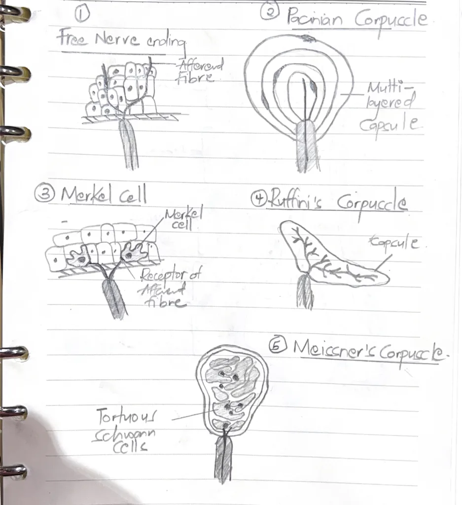
Describe the structure and function of the neural elements in skin
Unencapsulated nerve endings
Free nerve endings: fine touch, heat, cold, pain
Encapsulated nerve endings
Pacinian corpuscles: pressure changes and vibrationMeissner’s corpuscles: Sensitivity to light touchRuffini’s corpuscles: Sensitive to skin stretch and torque