Overview
Pelvimetry is the measurement of the bony dimensions of the birth canal to determine whether vaginal delivery is possible.
A pelvis with an adequate size and shape is required for vaginal delivery. The best indicator of an adequate pelvis is good progress during labour.
The adequacy of the pelvis can also be assessed using clinical pelvimetry. 3 planes are assessed: the inlet, mid-pelvis, and outlet.
Timing for clinical pelvimetry
| Category | Timing for clinical pelvimetry |
|---|---|
| Primigravida | Beyond 37 weeks |
| Multigravida | During labor |
Quick estimation of pelvic adequacy
| Measurment | Estimation |
|---|---|
| Diagnoal conjugate | Ability to touch the tip the sacral promontory |
| Inter-ischial diameter (transverse diameter of mid-pelvis) | Ability to touch the ischial promontory with two fingers simultaneously |
| Inter-tuberous diamter (transverse diameter of the outlet) | Ability to accomodate a fist between the ishcial tuberosities |
| Sub-pubic angle | Ability to place 2 fingers under the symphysis comfortably |
Pelvic Inlet
The pelvic inlet is also known as the pelvic brim. It is involved in fetal engagement. The fetal head enters the inlet in a transverse position since the transverse diameter of the inlet is wider than the anterior-posterior (AP) diameter.
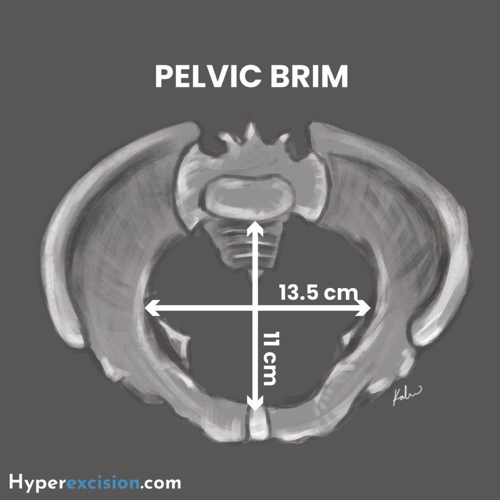
Boundaries of the pelvic inlet
| Boundary | Structure |
|---|---|
| Anterior | Upper border of the pubic symphysis |
| Lateral | Upper margins of the pubic bone, iliopectineal line, and ala of sacrum |
| Posterior | Sacral promontory |
Measurements of the pelvic inlet
| Measurement | Estimation |
|---|---|
| AP diameter (11cm) | Reach the middle finger to the sacral promontory and measure the distance to the pubic symphysis (diagonal conjugate). Subtract 1.5 cm to get the obstetric conjugate |
| Transverse diameter (13.5 cm) | |
| Pelvic shape | Slide fingers along the pelvic brim to assess contour (gynaecoid vs android) |
| Retropubic angle | Palpate behind the pubic symphysis. Flat and narrow suggest non-gynaecoid pelvis |
Mid-pelvis
The mid-pelvis is also known as the mid-cavity. The transverse and AP diameters of the mid-pelvis are almost equal at 12 cm.
Boundaries of the mid-pelvis
| Boundary | Structure |
|---|---|
| Anterior | Middle of pubic symphysis |
| Lateral | Pubic bones, obturator fascia, and inner aspect of ischial bones and spine |
| Posterior | Junction of the sendon and third section of the sacrum |
Measurements of the mid pelvis
| Measurement | Estimation |
|---|---|
| Ischial spines | Palpate for prominence or bluntness |
| Lateral pelvic sidewalls | Run fingers along the sidewaal for diverging (gynecoid) or converging (android) side-walls |
| Sacral curve | Deep J-shaped sacral curve is favourable for internal rotation while a flat sacrum is unfavourable |
Pelvic Outlet
The pelvic outlet provides space for delivery. The AP diameter is widest at the outlet, which means that the fetal head must rotate from a transverse to an AP position as it passes through the mid-pelvis.
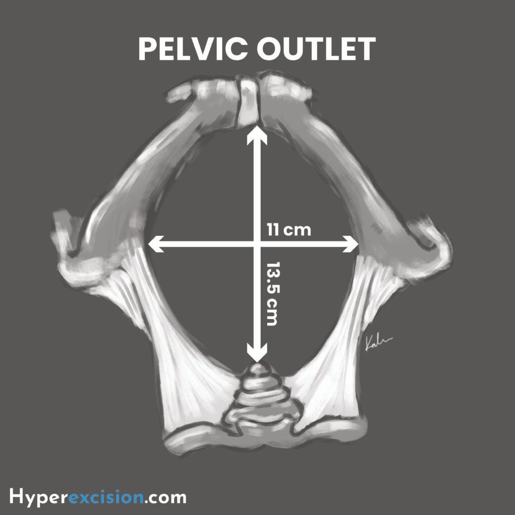
Boundaries of the pelvic outlet
| Boundary | Structure |
|---|---|
| Anterior | Lower margin of the symphysis pubis |
| Lateral | Inferior ramus of the pubis, ischial tuberosity, and sacrotuberous ligament |
| Posterior | Distal sacrum |
Measurments of the pelvic outlet
| Measurement | Estimation |
|---|---|
| Sub-pubic angle | A wide angle of 90 – 100 is favourable |
| A-P diameter (13.5 cm) | Distance from the pubic symphysis to the lower body of the pubic symphysis |
| Interuberous diameter | Fit a closed fist between the ischial tuberosities of the perineum |
Contracted Pelvis
A contracted pelvis is rarely discovered in clinical practice. Anatomically, it is defined as any of its major diameters that is shortened by 0.5 cm or more.
Thomas dictum: if the sum of the bispinous diameter and the posterior sagittal diameter of the outlet is < 15 cm, then the pelvis is likely contracted
Diagnostic criteria
| Contracted pelvis | Definition |
|---|---|
| Contracted inlet | Obstetric conjugate < 10 cm; Diagonal conjugate < 11.5 cm |
| Contracted mid-pelvis | Inter-ischial diameter < 8 cm |
| Contracted outlet | Intertuberous diameter < 8 cm |
Grossly abnormal pelvic types
| Pelvis | Description |
|---|---|
| Rachitic pelvis | Pelvic deformity due to childhood rickets |
| Robert’s pelvis | Bilateral absence of pelvic alae |
| Naegele’s pelvis | Unilateral absence of a pelvic ala |
Diameters of the Fetal Skull
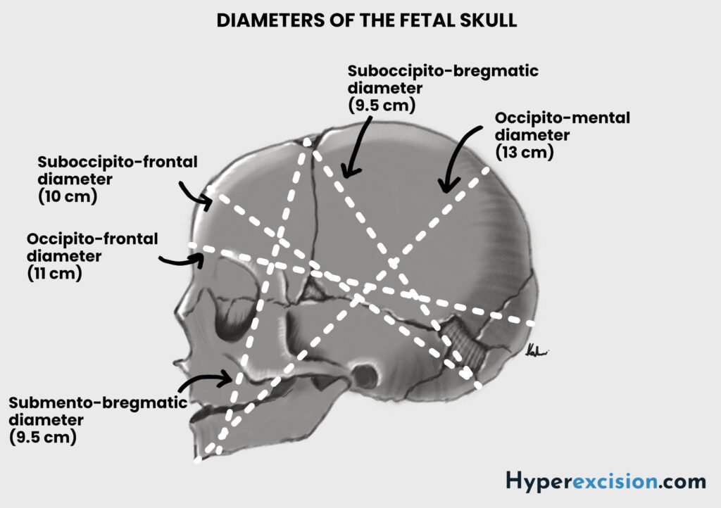
The fetal skull is ovoid in shape and is composed of sutures that allow the bones to move together and overlap. The parietal bones usually slide over the frontal and occipital bones, a process known as moulding, which reduces the diameters of the fetal head and encourages progress through the pelvis while protecting the underlying brain. Severe moulding or moulding in early labour can be a sign of obstructed labour due to cephalopelvic disproportion or malposition (failure of the head to rotate).
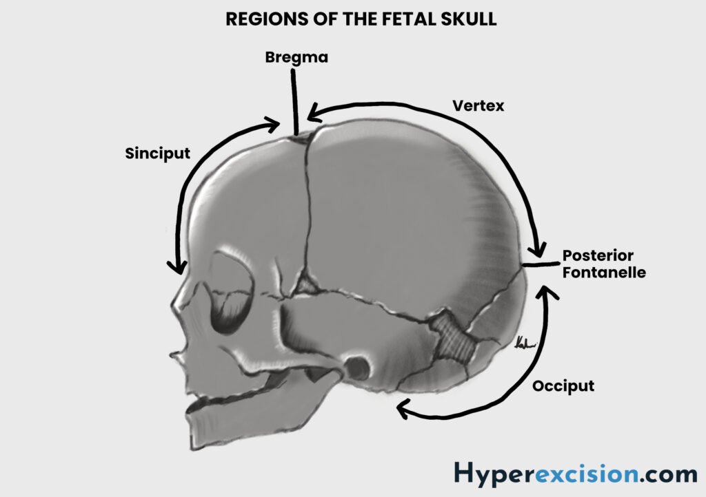
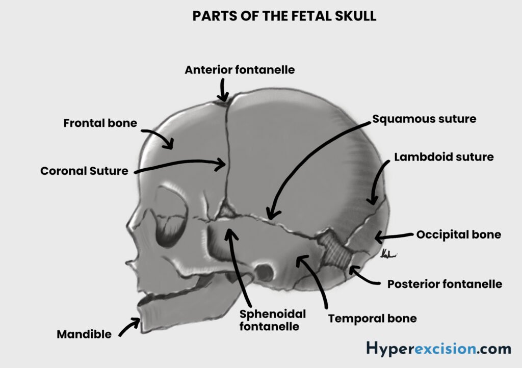
Transverse diameters of the fetal skull
| Diameter | Measurement | Nota bene |
|---|---|---|
| Biparietal | 9.5. cm | Engagement occurs when biparietal diameter passes the pelvic inlet i.e. it is at 0 station. Moulding can reduce biparietal diameter slightly (~ 0.5 cm) as the parietal bones overlap |
| Bitemporal | 8 cm | Narrower than biparietal diameter |
| Bimastoid | 7.5 cm | Smallest transverse diameter |
Longitudinal diameters of the fetal skull
| Diameter | Measurment | Attitude | Nota bene |
|---|---|---|---|
| Suboccipito-bregmatic | 9.5 cm | Well flexed | Smallest presenting diameter in a fully flexed vertex presentation |
| Occipito-frontal | 11.5. cm | Deflexed (Military presentaiton) | Increased risk of obstructed labour |
| Mento-vertical | 13.0 cm | Extended (Brow-presentation) | Incompatible with pelvic inlet. Requires caesarean delivery |
| Submento-bregmatic | 9.5. cm | Hyperextended (Face-presentation) | Face-presentation can deliver vaginally when the chin is anterior (mento-anterior) since this is the smallest diameter. Most meto-posterior presentations convert to mentum anterior |