History
A 55-year-old post-menopausal woman presents with a new breast mass in her right breast.
Q1: What is the significance of her age in this case?
She is post-menopausal. Breast carcinoma can occur at any age but frequency increases at 40 years. A new breast mass in a post-menopausal woman is cancer unless stated otherwise. She is less likely to be affected by masses that affect premenopausal women such as fibroadenoma and fibrocystic disease.
Q2: For her history of the presenting complaint, what information would you like to know about the new breast mass?
- Location of the mass
- Since when has the mass been present
- Rate of growth: rapid vs gradual
- Is the mass painful?
- Is the mass ulcerated?
- Are there any other skin changes?
- Does the mass shift position within the breast?
- Is there nipple retraction?
- Is there nipple discharge?
- Is there a mass in the opposite breast?
- Common causes of breast pain include: the luteal phase of the menstrual cycle, pregnancy, hematoma, cysts, mastitis, abscess, galactocele, and nipple fissure Malignant masses are commonly painless Hard fibroadenoma AKA breast mouse is commonly associated with shifting position Swelling found in both breasts include fibroadenoma, fibrocystic change and 3% of breast carcinoma
She states that the mass has been there for about 3 months and has slowly grown in size. She first noticed it when she was taking a shower. The mass is not painful. She reports no nipple discharge, no nipple inversion, and no skin changes.
Q3: What questions would you ask her to determine the likely cause of the breast mass?
- Her Last Normal Menstrual Period
- Her First Menstrual Period
- Her Parity and Last Delivery
- Her breastfeeding History
- History of Oral Contraceptive Use or Hormonal Replacement Therapy
- History of Breast or Ovarian Malignancy in her sister or mother
- History of trauma to the breast
- History of breast cancer
All breast masses that persist through one complete menstrual cycle, and all masses in post-menopausal women require evaluation. Remember that a normal mammogram or non-visualization of a palpable mass by ultrasound does not exclude cancer
There is a 5x risk for breast carcinoma with a family history of breast or ovarian cancer. Additionally, there is a very high risk of BRCA1 and BRCA2 mutations
Nulliparous women and women who do not breastfeed have an increased risk of breast cancer
She had her first menstrual period at 11 years of age. Her only pregnancy was at 35 years of age. Her mother and sister both had breast cancer.
Q4: What additional symptoms do you need to rule out that would suggest that the breast mass is “complicated”?
- History of Fever
- History of Purulent Discharge
- History of weight loss
- History of swelling in the armpits or neck
- History of shoulder pain, back pain, or abdominal swelling
- History of coughing up blood or breathlessness
- History of yellow discoloration of the eyes
- History of weakness, impaired sensation, headache, or blurry vision
- Fever, pain, and purulent discharge would indicate an infective process
Physical Examination
You proceed to perform a physical examination, starting with an inspection of the breasts.
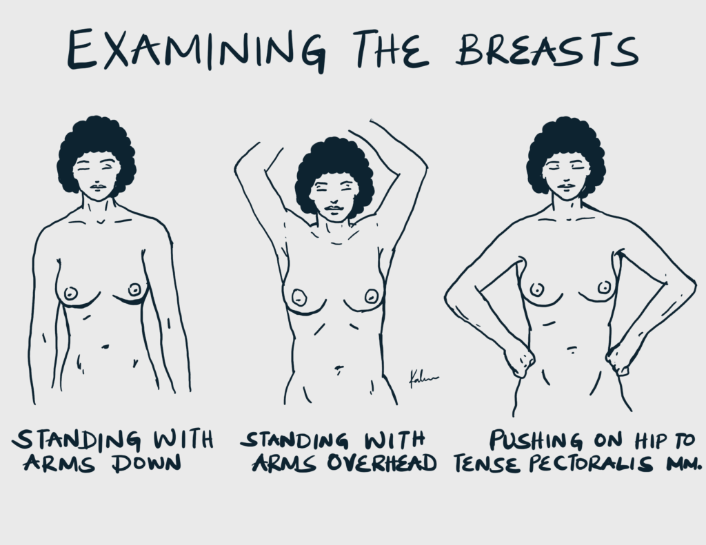
Q5: How should the patient be positioned to properly inspect the breasts?
- Sitting with hands on the hips
- Sitting with hands overhead
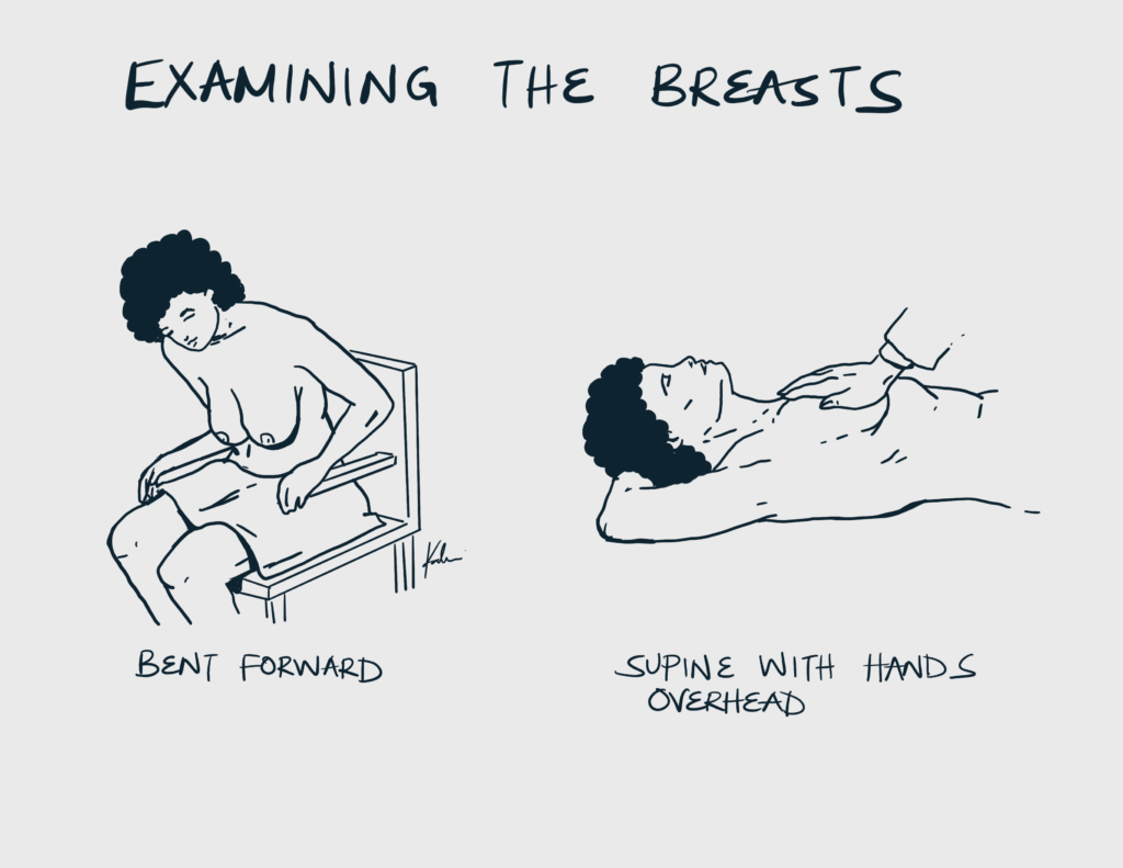
- Supine with hands overhead
The best time to examine the breast is 5 to 7 days after the onset of menses. The breasts may be engorged before menses, and during pregnancy making examination more painful and less accurate.
Both breasts are examined sequentially both sitting and supine
Pressing the hands on the hips may bring out dimpling by putting tension on the breast ligaments arising from the pectoralis major fascia
Raising the hands overhead stretches the ligaments of Cooper causing the skin or nipples to retract. It also allows us to expose the undersurface of the breast and examine the axilla
Q6: What findings are you looking for on inspection of the breasts?
Inspection of the breast
- Compare breast position
- Obvious mass
- Visible swelling in the axilla or neck
- Swelling of the arms
Swelling of the arms may due to lymphatic spread, post-operative axillary vein thrombosis or lymphedema following radical lymphadenectomy of the axillary nodes
Inspection of the skin over the breast
- Ulceration
- Dimpling or retraction
- Peau d’ orange
- Dilated veins
Ulceration is seen in a fungating carcinoma, cystosarcoma phyllodes and a rapidly growing fibroadenoma due to pressure atrophy
Try to pass a probe beneath the skin. It is possible to do so in benign conditions but impossible in malignancy
Peau d’Orange is a pitted appearance caused by cutaneous edema. The pitted marks are attachments or openings of follicles and sweat glands where the skin is firmly attached. It is seen in inflammatory carcinoma due to blockage of the lymphatic ducts.
Dilated veins may indicate Mondor’s disease, a huge abscess, phyllodes tumor or sarcoma
Inspection of the nipples
- The presence or absence of nipples
- Position
- Number
- Retraction
- Cracks and Fissures
- Discharge
Circumferential retraction may indicate breast cancer – the tumor infiltrates and shortens major ducts causing retraction Slit-like retraction may indicate duct ectasia w/ periductal mastitis Bilateral nipple retraction may be congenital Unilateral nipple retraction may indicate carcinoma or may follow fibrosis of a chronic abscess For discharge ask the patient to express. Also check whether retraction reverts as the attempt to express.
Inspection of the Areola
- Redness
- Shininess
- Oedema
Differentiate between eczema and Paget’s disease when examining the Areola Paget’s disease is Unilateral and occurs commonly in older women, there is no itching and vesicles and there may be destruction of the nipple and underlying lump Eczema is bilateral and occurs commonly in younger women. There is itching and vesicles. There is no destruction of the nipple and underlying lump. Unlike Paget’s disease, it has a quick response to local steroids
Q7: How should the patient be positioned to properly palpate the breast?
Bimanual palpation should be performed with the patient lying supine with the ipsilateral arm raised above her head
Q8: What findings are you looking for while palpating the breasts?
Palpation of both breasts
- Locate the mass
- Warmth
- Tenderness
Palpation of the mass
- Number of masses
- Location
- Size in 3 dimensions
- Shape
- Margins
- Consistency
- Fluctuation
- Tenderness
- Fixity to skin
- Fixity to breast tissue
- Fixity to chest wall
Mass with irregular surface and ill-defined margins may indicate breast carcinoma
Mass with well-defined margins and globular in shape are benign processes such as breast cysts and fibroadenoma
A cyst is a collection of fluid in a cavity lined by epithelium or endothelium
Breast cysts are fluctuant. They can also occur in fibroadenomas, cystic degeneration in malignancy, chronic abscesses
Fibromas have a firm consistency
Fibroadenomas are hard and feel like a pellet (diffuse shotty feel)
Carcinomas are usually hard to stony hard
Cysts, lipomas, and abscesses are soft in consistency
For fixity, pinch the skin over the mass and see whether it moves. Breast cancer, traumatic fat necrosis, and chronic abscesses are usually firmly tethered to the skin
Palpation of the axilla
- Lymphadenopathy
- Are the lymph nodes discrete or matted?
- Are there fixed or mobile lymph nodes?
- Is there tenderness?
- What is the consistency of the lymph nodes?
Group I nodes: Pectoral, Subscapular, Brachial
Group II nodes: Interpectoral, Central
Group III nodes: apical and supraclavicular
With the shoulder adducted, forearm flexed and resting over your forearm
Push fingers as high as possible to palpate the central nodes
Run fingers against the anterior axillary fold to palpate pectoral nodes
Run fingers against the humerus to palpate the brachial nodes
Stand behind the patient and run fingers against the posterior axillary fold to palpate the subscapular node
Then palpate the deltopectoral grove and superior to the middle 1/3 of the clavicle
On physical examination, she has a 2cm palpable, hard, ill-defined, immobile, non-tender mass in the upper outer quadrant of her right breast. There is no palpable axillary or supraclavicular lymphadenopathy.
Q9: What are the differentials for a benign palpable breast mass?
- Fibrocystic change: lumpy breast, upper quadrant, blue-dome appearance of cysts in physical exam
- Fibroadenoma: well-circumscribed mobile, rubbery, encapsulated mass
- Intraductal papilloma: unilateral blood nipple discharge in premenopausal women
- Fat necrosis: trauma to the breast or breast surgery, accompanied by pain
- Abscess: painful mass in lactating breast, erythematous and warm, fever, purulent discharge from the mass or nipple
- Galactocele: painful or painless aseptic mass in lactating breast, neither warm nor erythematous
Q10: What are the differentials for a malignant palpable breast mass?
- Ductal carcinoma in situ: not palpable
- Invasive ductal carcinoma: firm, immobile, discrete mass, nipple retraction, painless
- Invasive lobular carcinoma: firm, immobile, discrete mass, nipple retraction, painless, frequently bilateral
- Mucinous carcinoma: gelatinous well-circumscribed mass
- Inflammatory carcinoma: inflamed, tender, warm, erythematous breast, peau d’orange
- Phyllodes tumor: mobile, slow-growing, firm, rubber, and large
Q11: What is the most likely diagnosis for this woman?
- Invasive Breast Cancer
- Post-menopausal woman with new palpable breast mass
- Her risk factors for breast cancer include a family history in a first-degree relative and early menarche
- The mass is non-tender, hard, ill-defined, immobile, and in the upper outer quadrant
Investigations
Q12: What investigations would you perform to confirm the diagnosis, grade, and classify the tumor?
- Mammography
- Core needle biopsy
This is part of the triple assessment of breast cancer – physical examination, imaging, tissue sample for pathology
Imaging is done to rule out malignancy, and multicentricity and to screen women older than 30 years
Mammography is generally for women > 35 years and ultrasound < 35 years due to denser breast tissue. Ultrasound can also be used for needle-guided aspiration
FNAC is performed for cystic masses that are painful or enlarging
MRI is not indicated for the workup of a new breast mass due to its high false-positive rate
Q13: What imaging findings on mammography are suspicious of malignancy?
- Asymmetry
- Clustered pleomorphic calcifications
- Increasing density
- New mass with irregular borders or spiculations
Q14: What investigations would you perform following the diagnosis of breast cancer?
Metastatic workup
- Liver Function Test: for liver metastasis
- Alkaline phosphatase and serum calcium: for bone metastasis
- Chest X-ray: for pulmonary metastasis
Further Investigations for metastasis
- Abdominal and Chest CT: if abnormal chest X-ray and laboratory values
- Bone scintigraphy: suspicious bone pain or abnormal bone studies
- Brain CT or MRI: new onset headache, visual changes, or seizures
- PET scan
Treatment
Q15: What surgical options are available for a patient with Stage I and II Breast cancer
- Breast-Conserving Therapy (BCT): Lumpectomy (Partial Mastectomy) + Sentinel Lymph Node Biopsy (SLNB; 2-4 nodes) + Radiation (to decrease risk of local recurrence)
- Equal survival to mastectomy
- High local recurrence rate
- Simple Mastectomy with SLNB only
- Radiation is not needed in early stage
Q16: What are the surgical options for clinically advanced stage 3 breast cancer
- Simple mastectomy: if a large primary tumor
- Simple mastectomy with axillary lymph node dissection (SLND): if nodes are enlarged and matted
- Modified radical mastectomy (Mastectomy + ALND)
- Neoadjuvant chemotherapy followed by Modified radical mastectomy, then irradiation: for inflammatory breast cancer
Q17: What are the contraindications to BCT
Absolute contraindication
- Multiple primary tumors in 2 or more quadrants
- Diffuse malignant microcalcifications throughout the breast
- History of chest wall irradiation
- Positive margins despite repeat excision
- Early pregnancy (radiation cannot be given during pregnancy)
Q18: What are the options for hormonal therapy
- Estrogen Receptor Blockers: Tamoxifen, Raloxifene, Fulvestrant
- Aromatase inhibitors: Anastrozole
- HER2/Neu Receptor blocker: Trastuzumab
Q19: What drug classes are used for chemotherapy in breast cancer?
- Anthracyclines: Doxorubicin, epirubicin, idarubicin
- Alkylating agent: Cyclophosphamide
- Antimetabolites: Methotrexate, 5-FU
- Taxanes: Paclitaxel, Docetaxel
Q20: Which patients do not need chemotherapy?
- Carcinoma in situ
- Small (<1 cm) tumor with favorable hormonal and molecular characteristics
- SLNB negative
Complications
Q21: What nerves are at risk of damage during dissection
- Long thoracic
- Thoracodorsal
- Medial pectoral
- Lateral pectoral
Q22: What is the most severe complication of lymph node dissection
- Lymphedema
- ALND is associated with a greater risk of lymphedema than SLNB
- Lymphangiosarcoma in chronic cases (Stuart Treves syndrome)
- Has a poor prognosis even after limb amputation
This is why SLNB is very useful. It prevents unnecessary ALND
Key Learning Points
- New breast masses in post-menopausal women should be considered malignant until proven otherwise.
- Thorough history should include mass characteristics, associated symptoms, menstrual/reproductive history, and family history of breast/ovarian cancer.
- Proper physical examination technique is crucial, including inspection and palpation in different patient positions.
- The triple assessment for breast masses includes physical examination, imaging, and tissue sampling.
- Differentiate between benign and malignant causes of breast masses based on clinical features.
- Treatment options vary based on cancer stage, ranging from breast-conserving therapy to mastectomy with lymph node management.
- Consider contraindications to breast-conserving therapy when planning treatment.
- Be aware of potential surgical complications, especially lymphedema following axillary lymph node dissection.
- Adjuvant therapies include radiation, hormonal therapy, and chemotherapy, with selection based on tumor characteristics and stage.
- Sentinel lymph node biopsy can help avoid unnecessary axillary lymph node dissection in early-stage cancers.