A 32-year-old man is brought to the emergency department after a motorbike accident. He reports severe pain in the left knee and inability to bear weight. On inspection, there is gross swelling and deformity of the knee joint. The leg appears shortened and displaced laterally. He is in severe distress. Distal pulses in the dorsalis pedis and posterior tibial arteries are absent. The foot is pale and cool to the touch. Sensation over the dorsum of the foot is reduced, and he is unable to dorsiflex the ankle.
Q1. What is the most likely diagnosis?
Posterior knee dislocation with neurovascular injury
Knee dislocation is a true orthopaedic EMERGENCY
It occurs when there is rupture of at least two of the four major knee ligament structures. They can be due to a high-energy mechanism (50%), a sports injury (33%), or a low-energy mechanism (such as a fall).
Dislocations due to high-energy mechanisms have a higher risk of vascular injury
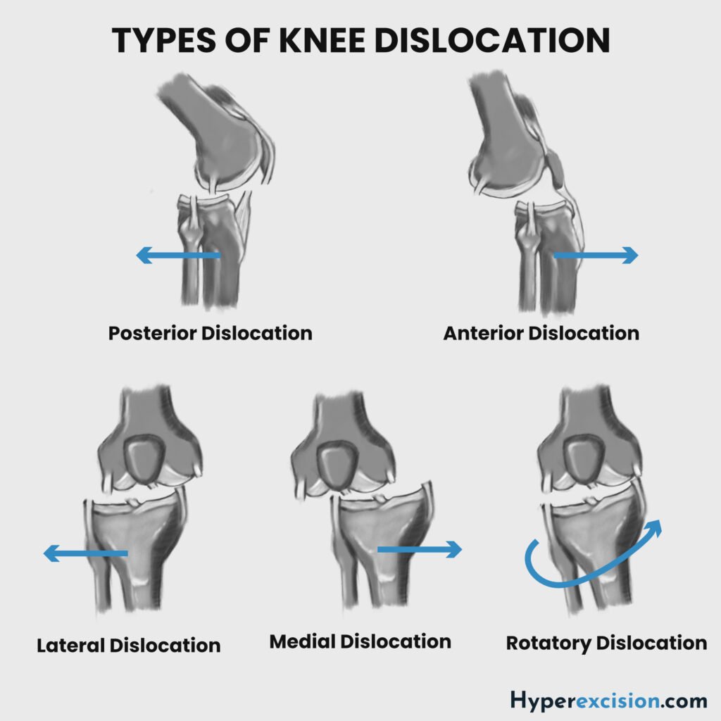
Q2. Classify knee dislocations (based on the direction of displacement of the tibia)
| Classification | Description |
|---|---|
| Anterior dislocation (40%) | Direct blow to the proximal tibia (dashboard injury). High risk of vascular injury due to disruption of the popliteal artery |
| Posterior dislocation | Direct blow to the proximal tibia (dashboard injury). High risk of vascular injury due to disruption of the popliteal artery |
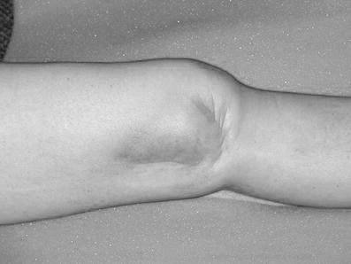
| Medial dislocation | Rare and usually irreducible. The PCL remains intact as the tibia rotates around the femur. It presents with a dimple on the medial side. Associated with the “dimple” sign. |
| Lateral dislocation | Due to a varus force |
| Posterolateral (Rotatory) dislocation | Rare and usually irreducible. The PCL remains intact as the tibia rotates around the femur. It presents with a dimple on the medial side. Associated with the “dimple” sign. |
Q3. What are the typical physical exam findings in a knee dislocation?
Overweight or obese (risk factor)
Hyperextension of the knee
Deformity
Knee swelling
Popliteal ecchymosis
Dimple sign (rotatory dislocation, may be irreducible)
Neurovascular examination
Capillary refill, skin color, and temperature
Distal pulses (dorsalis pedis and posterior tibial)
Ankle-brachial index (ABI)
Foot drop (L5 – common peroneal nerve injury)
Q4. What are the hard signs of limb ischaemia?
Absent pulse
Bleeding
Expanding hematoma
Cold cyanotic foot
Require urgent surgery for exploration (vascular surgery)
Soft signs include weak pulse, prolonged capillary refill, colour or temperature changes
Q5. What is included in the acute management of a knee dislocation?
Resuscitation
Consent for closed reduction
Analgesia and sedation
Pre-reduction neurovascular examination
Closed reduction
Post-reduction neurovascular examination and ankle brachial index
Plaster immobilization or external fixator (if unstable or vascular repair needed)
Non-weight bearing with crutches
Rotatory dislocation requires open reduction via an anteromedial approach after neurovascular examination
A temporary external fixator is used for unstable patients, persistent subluxation, or severe soft tissue injury
If there is a fracture dislocation, reduce the fracture, then a post-reduction CT scan is obtained. MRI is ordered too, before definitive repair.
Q6. What are the possible post-reduction ankle-brachial index (ABI)/neurovascular exam findings following closed reduction of a knee dislocation?
| ABI and neurovascular exam | Intervention |
|---|---|
| > 0.9 | Observation with serial neurovascular examinations over 24 – 48 hours |
| < 0.9 with asymmetric pulses | CT angiography or arteriography to evaluate the blood vessels |
| Absent pulses or clear hard signs of ischaemia | Emergency surgical exploration (vascular surgery). If ischaemia > 6 hours → urgent revascularization +/- fasciotomy |