Last updated:
November 12, 2024
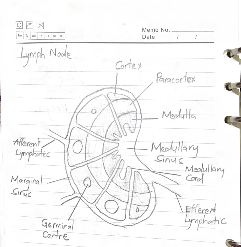
- Describe the general architecture of the lymph node
- Parenchyma
- Cortex: Outer portion of the lymph node except the hilum
- Superficial (Nodular) cortexDeep cortex (Paracortex): Contains most of the T-cells (Thymus-dependent)
- Medullar sinus: Opening that drains at the hilum into efferent vesselsMedullary cords: Contain reticular fibers, B lymphocytes, Macrophages, Dendritic cells, and plasma cells
- Cortex: Outer portion of the lymph node except the hilum
- Parenchyma
- Describe the events and cells involved in ‘thymic education
- Double-negative stage: T-cells do not yet express CD4 and CD8 molecules on their surface
- Double-positive stage: As maturation continues, T-cells express CD4 and CD8 molecules on their surface
- Type II and Type III epithelioreticular cells present T-cells with foreign and self antigens
- Positive selections: Occurs in the cortex. Cells that recognize self-MHC and Self/Foreign antigens progress to the medulla: where they undergo negative selection
- Negative selection: Cells that recognize self-MHC and self-Antigens are eliminated
- Single-positive stage: Cells that survive negative selection either become Cytotoxic CD8+ T-cells or Helper CD4+ T-cells (By stopping expression of either CD8 and CD4 molecules on their surface)
- What are Hassall’s corpuscles
- Hassal’s corpuscles are isolated concentric arrangements of type IV epithelioreticular cells. These cells contain keratohyalin granules, and are joined by desmosomes; the centers show keratinization.
- Their function is to produce interleukines (IL-4 and IL-7) that are necessary for the differentiation and education of T-cells.
- Briefly describe the open and closed circulation of the spleen
- Open circulation: Pencillar arterioles empty directly into the cords of Billroth rather than connecting to the endothelium-lined splenic sinuses. Blood entering the red-pulp seeps through the cords and is exposed to macrophages residing there.
- Closed circulation: Pencillar arterioles empty directly into the splenic sinuses. The endothelium of the pencillar arterioles and that of the splenic sinuses is continuous.