Overview
Gestational trophoblastic disease is the presence of abnormal tissue derived from fetal cells. These are also known as molar pregnancies or hydatidiform moles.
hCG mimics TSH, LH and FSH
Classification of Gestational Trophoblastic Disease
| Classification | Example |
|---|---|
| Benign (75%) | Complete (90%) and incomplete (10%) molar pregnancy |
| Malignant (25%) | Peristent or invasive mole, choriocarcinoma, placental site trophoblastic tumor (PSTT) |
hCG levels in Pregnancy and Time for Normalization
hCG levels peek around 9 – 12 weeks before falling.
Normal hCG range during pregnancy
| Weeks LMP | hCG levels (mIU/ml) |
|---|---|
| 3 weeks | 5 – 50 |
| 4 weeks | 4 – 426 |
| 5 weeks | 18 – 7340 |
| 6 weeks | 1080 – 56500 |
| 7 – 8 weeks | 7650 – 22900 |
| 9 – 12 weeks | 25700 – 28800 |
| 13 – 16 weeks | 13300 – 25400 |
| 17 – 24 weeks | 4060 – 165400 |
| 25 – 40 week | 3640 – 11700 |
Time for normalization
| Pregnancy | Normalization |
|---|---|
| Normal pregnancy | 4 weeks |
| Partial mole | 8 weeks |
| Complete mole | 14 weeks |
Molar Pregnancy
A molar pregnancy is an abnormal pregnancy that results from chromosomal irregularities. Early pregnancy bleeding is the most common presenting symptom and bleeding may include passage of hydropic villi (”grape-like” mass). A partial mole is commonly confused with a missed abortion (os is closed, no fetal heart rate). Blood should be on hand when evacuating molar pregnancies since this procedure is bloody.
| Complete mole | Partial mole | |
|---|---|---|
| Cause | Uniparental disomy – fertilizing an empty egg then duplication or dispermic fertilisation of an empty egg | Trisomy – dispermic fertilisation of an egg with maternal genetic material |
| Karyotype | 46XX, 46XY | 69XXY, 69XXX |
| Histology | Diffuse hydropic villi | Partial hydropic villi |
| p57 | Negative | Positive |
| Trophoblastic proliferation | Marked | Minimal |
| Fetal pole | Not present | May be present |
| Fetal cardiac activity | Absent | Absent |
| Amniotic fluid | Absent | May be present (oligohydramnios) |
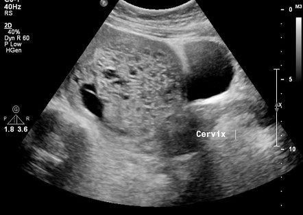
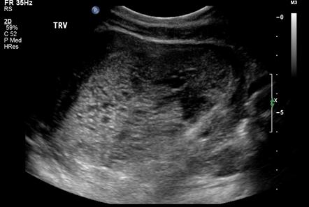
| Characteristic appearance | “Snowstorm appearance”- echogenic masses interspersed with hypoechogenic cystic spaces (hydropic villi) | “Swiss cheese” appearance – multicystic avascular hypoechoic or anechoic spaces |
| Malignant potential | Lower | Higher |
- Risk factors for molar pregnancy
- History of molar pregnancy (most important risk factor0
- Age ≤ 15 yo and ≥ 35 yo (extremes in age)
- History of miscarriage or infertility
- Blood group A with male partner Blood group O
- Vitamin A deficiency
- History of OCP use
- Smoking
- Signs and symptoms of molar pregnancy
- First-trimester bleeding
- Passage of a “grape-like” mass or clots (hydropic villi)
- High amount of nausea and vomiting (due to very high B-hCG)
- Palpitations, sweating, tremors (symptoms that mimic thyrotoxicosis due to B-hCG sharing a-subunit with TSH)
- Irritability, dizziness and photophobia (pre-eclamptic like symptoms – pre-eclampsia < 20 weeks is pathognomic for molar pregnancy)
- Uterus size abnormal for gestational age (28%)
- complete mole → large
- partial mole → small
- Adnexal masses may be felt (theca-lutein cyst due elevated LH and FSH activity)
- Investigations
- Transvaginal ultrasound
- Complete mole: prominent villi, snowstorm appearance, no fetus visible
- Partial mole: may show fetal pole
- Chest X-Ray: invasive moles can metastasize to the lungs
- Serial quantitive B-hCG: until it normalizes. B-hCG acts like a tumor marker. If it does not normalize we’re now dealing with gestational trophoblastic disease
- Pre-operative investigations: CBC, UEC, Coagulation panel, TFTs, group and cross-match (very bloody procedure)
- Transvaginal ultrasound
- Treatment of molar pregnancy
- Suction Dilatation and Curretage + Histology of last suction
- Serial B-hCG monitoring
- Contraception during Serial B-hCG monitoring
- RhoGAM if Rh negative
- Follow up after definitive management of molar pregnancy
- Sereial B-hCG levels weekly for 3 consecutive weeks until levels are normal (should fall in the 3 measurements)
- Follow-up monthly for next 6 months until level is zero
- Barrier contraceptives until hCG normalizes. Then hormonal can be started until normalized for at least 6 months
- Indications for prophylactic chemotherapy (Methotrexate 15 mg OD for 5 days )
- B-hCG > 100,000
- 6 cm lutein cyst
- 40 years
- High parity
- hCG takes about 2-4 weeks to clear after evacuation. What are the differentials for persistently elevated hCG levels after evacuation?
- Gestational Trophoblastic Disease (GTD)
- Retained Products of Conception (RPOCs)
- Another pregnancy
- What are the benefits of giving oxytocin during evacuation for molar pregnancies?
- Reduces bleeding (but can cause seeding)
- Reduced risk of uterine perforation
- Why should estrogen be avoided in case of suspected molar pregnancy?
- To prevent the risk of transforming to choriocarcinoma
- Complications of molar pregnancy
- Pre-eclampsia
- Hyperthyroidism
- Hyperemesis gravidarum
- Theca-lutein cysts
- Respiratory distress
- Hypovolemic shock
- Development of Choriocarcinoma
Malignant Gestational Trophoblastic Disease
50% of malignant GTD occur months to a year after a mola pregnancy. 25% occur after miscarriage or ectopic pregnancy. 25% occur after a normal pregnancy. Choriocarcinomas are more likely to follow a normal pregnancy while invasive GTD are more likely to follow a non-molar pregnancy.
Staging of GTD
| Stage | Extent |
|---|---|
| Stage I | Confined to uterus |
| Stage II | Metastases to pelvis and vagina |
| Stage III | Metastasis to Lungs |
| Stage IV | Distant metastases to liver, brain etc. |
Types of Malignant GTDs
| Malignant GTD | Description |
|---|---|
| Persistent or invasive mole | Follows evacuation fo molar pregnancy. Invades the myometrium and can metastasize. Diagnosed when hCG plateaus on follow-up |
| Choriocarcinoma | Malignant placental tissue. May occur in nulliparous women (ovaries) and men (testes). Gestational-related is most common. These can metastasize |
| Placental site trophoblastic tumors (PSTT) | Malignat tumor derived from cytotophoblasts at the placental implantation site. Can invade the myometrium but rarely spread outside the uterus. Does not respond well to chemotherapy and carries poor prognosis if it metastasiez |
- Signs and symptoms
- Per vaginal bleeding
- Late postpartum bleeding (> 6 – 8 weeks post-partum) for gestational-related choriocarcinoma
- Chronic, persistent, irregular bleeding (weeks to years after pegnancy) for PSTT
- Metastatic symptoms
- Lungs: cough, dyspnoea, respiratory distress, hemoptysis
- CNS: headache, dizziness, papilloedema
- Vaginal mass that easily bleeds
- Uterine enlargment
- Bilateral theca lutein cysts (due to high hCG)
- Per vaginal bleeding
- Investigations
- Quantitative hCG level:
- Elevated hCG (in the thousands) for choriocarcinoma and persistent mole
- hCG plateau on follow-up
- < 100 mIU/mL in PSTT
- Pelvic ultrasound: best test to diagnose PSTT
- Vacularized uterine mass
- Less vascularized uterine mass for PSTT
- Chest X-ray: baseline on diagnosis to see if there are any metastases to the lungs
- Quantitative hCG level:
- Treatment
- Low-risk: Methotrexate
- High-risk: Methotrexate, actinomycin D and etoposide
- Hysterectomy for PSTT followed by chemotherapy 1 week later
- Follow-up
- Sereial B-hCG levels weekly for 3 consecutive weeks until levels are normal (should fall in the 3 measurements)
- Follow-up monthly for next 12 months until level is zero
- Barrier contraceptives until hCG normalizes. Then hormonal can be started until normalized for at least one year