Cervical incompetence
Cervical incompetence is the inability of the cervix to hold closed during pregnancy. When there is incompetence, the cervix spontaneously opens up leading to spontaneous expulsion of the fetus. Cervical incompetence leads to recurrent preterm birth or second trimester pregnancy lossess.
Incompetence presents as painless cervical dilation in the absence of uterine contractions and/or labor. It can be anatomic or due to functional incompetence. It is commonly caused by cervical tears due to precipitous labor or curretage. Tears occur more commonly on the lateral side giving a fish-mouth apperance on examination. Obstetric History will show preterm births with stepwise decreasing gestational age. Treatment is by permanent or temporary stitches which should be removed at 37 weeks gestation.
NOTE: Most cases of cervical insufficiency have unknown causes. A history of previous pregnancy terminations increases the chances for preterm delivery as well.
Causes of cervical insufficiency
| Classification | Causes |
|---|---|
| Congenital (rare) | Mullerian duct anomalies, in-utero exposure to diethylstilbestrol, collagen vascular disorders e.g. Ehlers-Danlos syndrome |
| Acuired | Cervical conization (excisional biopsy to diagnose cervical dysplasia), overenthusiastic dilation and curretage, fetal delivery before full cervical dilation (precipitous labour), traumatic vaginal delivery, operative vaginal delivery, obstructed labour, annular tear or perforation of the cervix, mechanical dilatation of the cervix for gynaecological procedures |
- Diagnosis of cervical insufficiency
- Bad Obstetric History: occurence of ≥ 2 adverse events during pregnancy
- Recurrent pregnancy losses (≥ 2 consecutive spontaneous miscarriages)
- Stillbirth
- Intrauterine growht restriction
- Preterm birth
- Congenital anomalies
- Neonatal death
- Classical clinical history of recurrent preterm births or uterine contractions
- Easy passage of Hegar’s No.8 cervical dilator in the non-pregnant state
- Ultrasound findings of reduced cervical length (< 3 cm) and internal os funnelling in the pregnant sate
- Bad Obstetric History: occurence of ≥ 2 adverse events during pregnancy
- Pathophysiology
- The main problem is a structural defect in the fibrous connective tissue of the cervix causing reduced tensile strength due to the causes listed above.
- Weak cervix cannot sustain the weight and expansion of amniotic sac (uterine cavity is filled at 14 weeks – second trimester)
- ROM occurs followed by bleeding or contractions
- Severe cases can present with painless fall of amniotic sac
- Signs and symptoms
- Painless cervical dilatation
- Prolapse and Ballooning of the membranes in the vagina
- Spontaneous expulsion of the fetus
- Some women may present with cramping, pelvic pressure, increased vaginal discharge and back pain
- Short cervix
- Patuloous cervix with or without closed inner os
- Anatomical deficiency on cervix
- Differentials
- Investigations
- Cervicogram
- Cervical length < 3cm
- Funnelling of internal os >1cm
- Y-shaped cervix
- V-shaped cervix
- U-shaped cervix
- Fetal Fibronectin – place a swab in the posterior vaginal fornix to collect the sample and measure fetal fibronectin. This is a fetal glycoprotein that promotes cellular adhesion at the utero-placental interface. Its breakdown and release into vaginal secretions indicates that the risk of preterm delivery is high.
- Cervicogram
- Treatment
- Cervical cerclage at 12-14 weeks
- MacDonald cerclage sub-mucosal purse string suture
- Shirodkar cerclage: sub-mucosal continous mersilene tape
- Tracehlorrhaphy: suturing tears in the cervix
- Progesterone supplementation: Intramuscular or Intravaginal Progesterone if indication for cervical cerclage is not met. Women with a prior history of preterm birth should receive supplementation at 16-24 weeks and continued up to 36 weeks of gestation.
- Cervical pessaries: prosthesis inserted into the vagina that displace the weight of the pregnant uterus away from teh cervix
- Cervical cerclage at 12-14 weeks
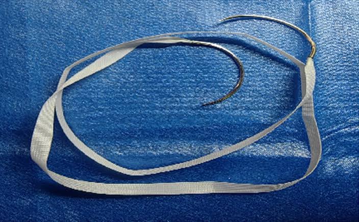
Cervical cerclage
Cervical cerclage is a surgical procedure used to reinforce the cervix in pregnant women with cervical incompetence. It usually invovles placing non-absorbable sutures (mersilene tape) around the cervix to provide mechanical support and prevent premature dilation of the cervix. Prophylactic stitches are usually placed at 12 – 14 weeks and removed by 36 – 37 weeks.
Types of cerclage
| Cerclage | Description |
|---|---|
| McDonald cerclage | Transvaginal purse-string suture placed sub-mucosal around the cervix at the cervicovaginal junction without bladder mobilization |
| Shirodkar cerclage | Transvaginal purse string sutures are placed submucosal higher in the cervix and require bladder mobilization to allow insertion above the cardinal ligaments. This is a permanent suture. |
| Transabdominal cerclage (TAC) | Permanent suture placed at the cervicoisthmic junction through laparoscopy or laparotomy when vaginal cerclage fails or is not feasible. Can be placed preconceptionally or in the first trimester |
- Indications for cerclage
- History-indicated cerclage (prophylaxis): placed at 12 – 14 weeks
- History of ≥ 2 second-trimester losses due to painless cervical dilatation
- Previous cerclage placement for cervical insufficiency
- Ultrasound-indicated cerclage
- Emergency cerclage
- Painless cervical dilation with prolapsed membranes in the second trimester (without infection or contractions)
- History-indicated cerclage (prophylaxis): placed at 12 – 14 weeks
- Contraindications for cerclage
- Pre-operative evaluation
- Ultrasound – for fetal viability, gestational age, rule out fetal abnormalities
- Clinical evaluation for active bleeding, and to rule out PPROM
- Amniocentesis to rule out infection
- Post-operative complications
- Chorioamnionitis
- Suture displacement
- Artificial rupture of membranes (AROM)
- Post-operative instructions
- Restric strenuous activity (on a case by case basia)
- Serial ultrasound for cervical length and signs of infection
- Return to remove cerclage at 36 – 37 weeks unless labour or complications occur earlier