History
A 17-year-old girl with a history of Type I Diabetes Mellitus is brought to the Emergency Department with nausea, vomiting, lethargy, and dehydration
Q1. What is the most likely diagnosis?
Reveal answer
Q2. What other symptoms are associated with DKA?
Reveal answer
- Abdominal pain
- Polyuria
- Polydipsia
Q3. What would be your top 3 differentials if this girl presented with acute onset abdominal pain, nausea, and vomiting?
Reveal answer
- Appendicitis
- Ectopic pregnancy
- Diabetic Ketoacidosis
Acute pancreatitis is also a differential!
Q4. What other endocrine emergency is associated with abdominal pain?
Reveal anser
Abdominal pain in both DKA and Addisonian crisis may be associated with Mesenteric ischemia.
The mother reports that she stopped taking her insulin a day before the presentation.
Q5. What are the precipitating factors for developing DKA?
Reveal answer
- Non-compliance
- Infections – Commonly UTI and Pneumonia
- Infarction – myocardial infarction and stroke
- Treatment failure +/- inability to afford treatment
- Surgery
- Trauma
- Burns
- Drugs e.g. Glucocorticoids, Cocaine
Anything that leads to an increased requirement of insulin can lead to DKA in patients with Type I Diabetes
Q6. What is the pathogenesis of DKA?
Reveal answer
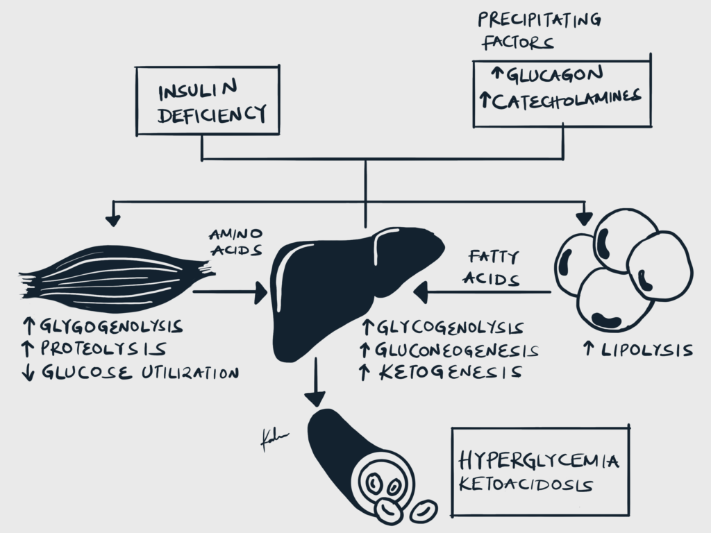
- Etiology
- DKA is commonly seen in T1DM, an autoimmune disease where the patients have complete/near total insulin deficiency. 70-80% of beta cells are destroyed in patients with T1DM and insulin is required for survival. Skipping a single day of insulin can have devastating consequences.
- Insulin deficiency and increased counter-regulatory hormones cause inhibition of glycolysis and increased gluconeogenesis and glycolysis → Hyperglycemia
- Insulin deficiency increases lipolysis through disinhibition of Hormone-sensitive lipase (HSL) → Lipolysis and generation of Free Fatty Acids → Ketogenesis
- Osmotic diuresis and hypovolemia
- Hyperglycemia causes hyperosmolality and osmotic diuresis which leads to volume depletion and hypovolemia
- Metabolic acidosis with increased anion gap
- Non-esterified fatty acids (NEFAs) generated from lipolysis are converted into ketones in the liver (acetoacetate, Beta-hydroxybutyrate)
- These ketones are acidic and bicarbonate is consumed as it buffers them.
- This causes metabolic acidosis with an elevated anion gap
- Intracellular potassium deficit
- Hyperglycemic hyperosmolality shifts potassium along with water from the intracellular to extracellular space
- Shifted potassium in the extracellular space is lost through urine
- Insulin is absent in DKA and can therefore not promote intracellular potassium uptake
- Total body potassium deficit develops although serum potassium may be normal or paradoxically elevated
Physical exam
She is a thin woman in mild respiratory distress. Respiratory rate is 28 breaths per minute, Blood Pressure 80/40mmHg, Heart Rate 112 beats per minute, Temperature 37.2 C. There are normal heart sounds, the lungs are clear, the abdomen is soft, and there is no organomegaly. She is responsive and oriented to time place, place, and person, but somnolent and weak. Mucous membranes are dry.
Q7. What signs are expected to be elicited on examination of a patient with DKA?
Reveal answer
- Ill-looking
- Signs of dehydration: dry skin, dry mucous membranes, decreased skin turgor
- Hypotension
- Tachycardia
- Tachypnoea (Kussmaul breatheing)
- Confusion
Q8. What homeostatic abnormalities are expected in DKA?
Reveal answer
- Hyperglycemia
- Acidemia
- Hypovolemia
- Depleted total body sodium, potassium and magnesium
- Acute Kidney Injury from volume depletion
Investigations
These are her lab results:
| Investigation | Value | Normal range |
|---|---|---|
| Serum sodium | 126 | 132 – 146 mEq/L |
| Potassium | 4.3 | 3.5 – 5.5 mEq/L |
| Magnesium | 1.2 | 1.3 – 2.1 mEq/L |
| BUN | 76 | 9-23 mg/dl |
| Creatinine | 2.2 | 0.5 – 1.1 mg/dl |
| Bicarbonate | 10 | 22 – 25 mmol/L |
| Chloride | 88 | 99 – 109 mEq/L |
| Serum glucose | 40 mmol/L (720 mg/dl) | < 11 mmol/L |
Q9. What are the 3 key diagnostic lab features of DKA?
Reveal answer
- Acidemia: Venous Blood pH <7.3 or HCO3- <15.0mmol
- Hyperglycemia: Blood Glucose >11mmol/L, Know Diabetic
- Ketonemia or significant ketonuria: ≥3.0mmol/L or 2+ on urinalysis respectively
Q10. Interpret these lab results
Reveal answer
- Hyperglycemia of 40 mmol/L. The degree of hyperglycemia does not correlate with severity. DKA can be seen with blood glucose ranging from “low” as 11 mmol/L to as high as 40 mmol/L as in this case.
- Pseudohyponatremia of 126 mEq/L. Corrected sodium (can be done using an online calculator) in this patient is 141 mEq/L which is within normal limit.
- Potassium of 4.3 mEq/L which is within the lower end. Concern for hypokalemia once treatment with insulin is started.
- Hypomagnesemia of 1.2 mEq/L which is common in DKA. Can be replaced with IV Magnesium.
- Low Bicarbonate of 10 mmol/L reflects metabolic acidosis (as bicarbonate is used as a buffer)
- Hypochloremia of 88 mEq/L from volume depletion resulting from vomiting and osmotic diuresis
- The anion gap is elevated and this is typical of DKA
Q11. What are the differentials for anion gap metabolic acidosis? (CAT MUDPILERS)
Reveal answer
- Carbon Monoxide, Cyanide
- Alcohol (Ethanol, Methanol)
- Toluene
- Uraemia
- DKA
- Propylene glycol, Paraldehyde, Paracetamol
- Iron tablets, Isoniazid
- Lactic acidosis
- Ethylene glycol
- Renal failure
- Salicylates, sepsis, starvation
Q12. What are the differentials for ketoacidosis?
Reveal answer
- Diabetic Ketoacidosis
- Alcoholic Ketoacidosis
- Starvation Ketoacidosis
Q13. What are the differentials for a patient with hyperglycemia and hypovolemia?
Treatment
Q14. What are the 3 prongs of managing DKA?
Reveal answer
- Potassium
- Intravenous Fluid
- Insulin
Q15. The management of DKA varies according to specific hospital policies. What are the general management steps of DKA?
Reveal answer
- Check blood glucose, ketone bodies, and potassium
- Administer IVF to replace fluid loss (usually NS at 2L in the first hour and 1L in subsequent hours) and Insulin
- Re-check blood glucose and ketone bodies
- If glucose is normal and ketones are high continue insulin and now give dextrose with potassium
- When glucose is normal and ketones are normal stop infusion and give potassium as needed
Potassium (K) is given before insulin, as insulin shifts K into cells and the large volume of IVF increases urine output and K wasting
IVF is given in large volume (LR = NS); if glucose lowers, but the anion gap doesn’t close, switch to a D5-containing solution
Insulin is given IV until the anion gap closes; once the anion gap is closed, switch to SQ insulin and start food (remember, in DKA the body thinks it is starving and activates starvation ketosis, which creates an anion gap)
Q16. What are the general fluid management guidelines for DKA?
Reveal answer
- 1-3 L of NS in the first hour
- 1 L in the second hour
- 1L next four hours
Q17. What is the general insulin guideline for managing DKA?
Reveal answer
IV Insulin at 0.1 u/kg/h
Q18. What is the role of insulin in DKA?
Reveal answer
- To drive glucose into cells
- To inhibit HSL and stop ketone body production
Q19. What is the hourly goal of insulin drop in DKA?
Reveal answer
- 4.4-5.5 mmol/L per hour
Hence, start the insulin drip at 0.1 U/kg/hr.
When glucose levels reach 13.9-16.6 mmol/L or it drops > 5.5 mmol/L/hr, add D5 to the fluids.
Once pH > 7.3, HCO3 > 16 mmol/L, anion gap is normal and the patient is eating, start SQ insulin and discontinue insulin drip after 1 hr.
Q20. When is insulin infusion stopped in a patient in DKA?
Reveal answer
- Once ketones return to normal and not when glucose levels are normal
Ketone bodies will still be produced even when glucose levels are normal. Insulin is needed for its inhibitor effects on HSL.
Q21. What are the general guidelines for replacing potassium in DKA?
Reveal answer
| Serum K+ | Amount of KCl to add per litre of IV fluid |
|---|---|
| >5.5 | Nil |
| 3.5-5.5 | 40 mmol |
| <3.5 | Stop insulin. Seek help from HDU/ICU for higher doses |
Complications and Prognosis
Q22. What is the best measure of severity in DKA?
Reveal answer
- Serum bicarbonate (anion gap) and not blood glucose
If serum bicarb is very low (very high anion gap) the patient is at risk of death.
| Severity | pH | Bicarbonate (mmol/L) |
|---|---|---|
| Severe | < 7.1 | < 5 |
| Moderate | 7.1-7.2 | 5-10 |
| Mild | 7.2-7.3 | 10-15 |
Q23. Why is bicarbonate only reserved for severe acidemia (pH < 6.9) in DKA?
Reveal answer
- Bicarbonate is converted into CO2 which causes widespread cerebral vasodilation and cerebral oedema.
- Bicarbonate increases the work of breathing and can exacerbate acidosis
Q24. What is the most feared complication of DKA?
Reveal answer
- Cerebral oedema
Especially in children when fluids are given too rapidly. Insulin bolus can also cause a sharp decrease in serum glucose and serum osmolarity.
Typically 10-20ml/kg NS boluses are given over 1 hour followed by repletion of the remaining fluid over 24-48 hours + maintenance fluid.
Key Learning Points
- History and Presentation
- Most Likely Diagnosis: DKA, especially with a history of Type 1 Diabetes Mellitus and symptoms like nausea, vomiting, lethargy, and dehydration.
- Associated Symptoms: Abdominal pain, polyuria, polydipsia.
- Differential Diagnoses for Acute Abdominal Pain, Nausea, and Vomiting: Appendicitis, Ectopic pregnancy, DKA, Acute pancreatitis
- Other Endocrine Emergency Associated with Abdominal Pain: Addisonian crisis (mesenteric ischemia might be involved).
- Pathogenesis of DKA
- Seen in Type 1 Diabetes Mellitus (T1DM) due to insulin deficiency.
- Insulin deficiency leads to hyperglycemia and ketogenesis.
- Hyperglycemia causes osmotic diuresis, leading to hypovolemia.
- Elevated anion gap metabolic acidosis due to acidic ketones.
- Intracellular potassium deficit despite normal or elevated serum potassium.
- Clinical Presentation
- Typical Signs: Ill appearance, dehydration, hypotension, tachycardia, tachypnea (Kussmaul breathing), confusion.
- Homeostatic Abnormalities: Hyperglycemia, acidemia, hypovolemia, depleted sodium and magnesium, potential acute kidney injury.
- Diagnosis
- Key Diagnostic Features: Acidemia (pH < 7.3 or HCO3- < 15 mmol/L), Hyperglycemia (Blood Glucose > 11 mmol/L), Ketonemia or ketonuria
- Differentials for Anion Gap Metabolic Acidosis include DKA, lactic acidosis, renal failure, and others.
- Differentials for Ketoacidosis include DKA, alcoholic ketoacidosis, starvation ketoacidosis.
- Differentials for Hyperglycemia and Hypovolemia include DKA, sepsis, acute pancreatitis.
- Management
- Three Prongs of Management: Potassium, Intravenous Fluids (IVF), Insulin.
- Monitor blood glucose, ketone bodies, and potassium.
- Administer IVF and insulin, adjusting as necessary.
- Initial bolus of 1-3 L of NS, followed by 1 L per hour.
- IV insulin at 0.1 U/kg/hr, adjust according to glucose levels and anion gap.
- Replace potassium before starting insulin to prevent hypokalemia.
- Stop Insulin once ketones are normal, not just when glucose levels normalize.
- Complications and prognosis
- Measure of Severity: Serum bicarbonate and anion gap, not just blood glucose.
- Bicarbonate Use: Reserved for severe acidemia (pH < 6.9) to avoid complications like cerebral edema.
- Most Feared Complication: Cerebral edema, especially in children, often due to rapid fluid replacement or insulin boluses.
References
Jameson, J.L. et al. (2018) Harrison’s principles of Internal Medicine. New York etc.: McGraw-Hill Education.