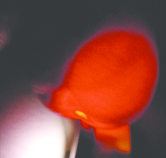
A 10-year-old male presents with a 2-month history of swelling of the left scrotum. The swelling has been increasing in size, but is not associated with pain, trauma, fever or urinary symptoms. On examination, the left hemiscrotum is enlarged, non-tender, smooth and fluctuant. It reduces in size when the patient lies down. The left testicle was palpated and felt to be normal and separated from the swelling. The swelling transilluminates in torchlight.
Q1. List the layers of the scrotum from superficial to deep
Reveal answer
Skin: highly elastic and loosley attached to underlying tissue
Tunica dartos: continuation of camper’s and scarpa’s fascia of the abdomen. contains the dartos muscle
External spermatic fascia: derived from external oblique aponeourosis
Cremasteric fascia and muscle: derived from the internal oblique and its fascia. Responsible for the cremasteric refle (elevates the testis)
Internal spermatic fascia: derived from the transversalis fascia
Tunica vaginalis: serous membrane derived from the peritoneum with a parietal and visceral layer. Below the tunica vaginalis is the testis which is enclosed by the tunica albuginea
Q2. What are the differentials for a painless scrotal mass in children?
Reveal answer
| Differential | Palpation | Transillumination | Increase with valsava |
|---|---|---|---|
| Tumour | Firm | No | No |
| Varicocele | ‘Bag of worms’ | No | Yes |
| Non-communicating hydrocele | Fluctuant | Yes | No |
| Communicating hydrocele | Fluctuant | Yes | Yes |
| Inguinal hernia (without incarceration) | Non-tender, reducible | No | Yes |
| Scrotal edema | Bilateral soft swelling | No | No |
| Spermatocele | Small, soft and localized cyst | Yes | No |
Q3. What is the diagnosis?
Reveal answer
Left-sided hydrocele
- Fluctuant, painless swelling
- Transilluminates in torchlight
Q4. What are the causes of this condition?
Reveal answer
Delayed closure of a patent processus vaginalis
Testicular torsion
Tumour
Epididymitis
Orchitis
Trauma
Hydroceles are common in newborns (due to delayed closure of the patent processus vaginalis or fluid trapped due to descent). The majority of hydroceles in neonates resolve spontaneously by 1 – 2 years of age.
Hydroceles in children and adolescents are usually reactive and must be evaluated to rule out secondary causes by palpation and/or ultrasound. They resolve once the underlying condition is treated.
Q5. How is it classified?
Reveal answer
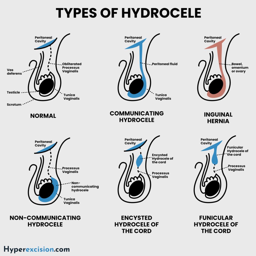
Classification of Hydroceles
| Classification | Characteristic | Examples |
|---|---|---|
| Communicating hydrocele | Patent processus vaginalis that communicates with the peritoneal cavity. Fluid can be “milked” into the peritoneum | Congenital, Funicular |
| Non-communicating hydrocele | No communicationbetween the processus vaginalis and peritoneal cavity | Vaginal, Infantile, Hydrocele of the cord |
| Hydrocele of the canal of Nuck | Non-tender inguinal-labial swelling in females due to failed closure of the canal of Nuck |
Communicating hydroceles develop due to failure of the processus vaginalis to close during development → fluid around the testis is peritoneal fluid
Non-communicating hydrocele is caused by fluid from the mesothelial lining of the tunica vaginalis.
Q6. What are the indications for surgical treatment of this condition?
Reveal answer
Does not resolve spontaneously by 1 year of age (to avoid the risk of indirect inguinal hernia and persistent enlargement)
Cosmetic concern
Pain and discomfort
Failure to reduce spontaneously after appropriate observation
Associated with suggestive pathology e.g. torsion or tumor
Pain or discomfort
Infertility is a concern i.e. due to infection, tumor, or injury etc.
Q7. What are the options for treatment?
Reveal answer
| Surgical procedure | Description |
|---|---|
| Herniotomy | For paediatric (communicating) hydroceles. Congenital hydroceles may be observed for 1- 2 years since they usually resolved on their own. |
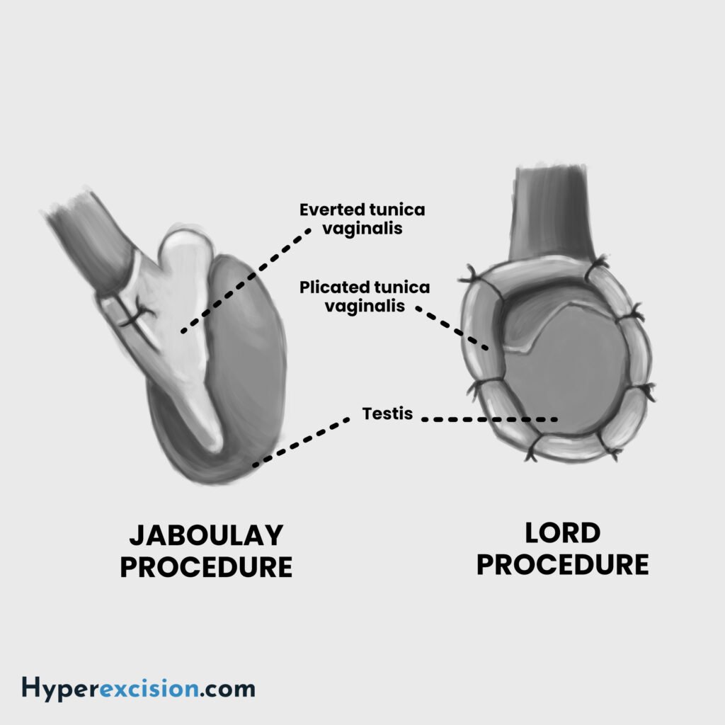
| Lord’s procedure | Plication of the tunica vaginalis |
| Jaboulay’s procedure | Excision and eversion of the sac |