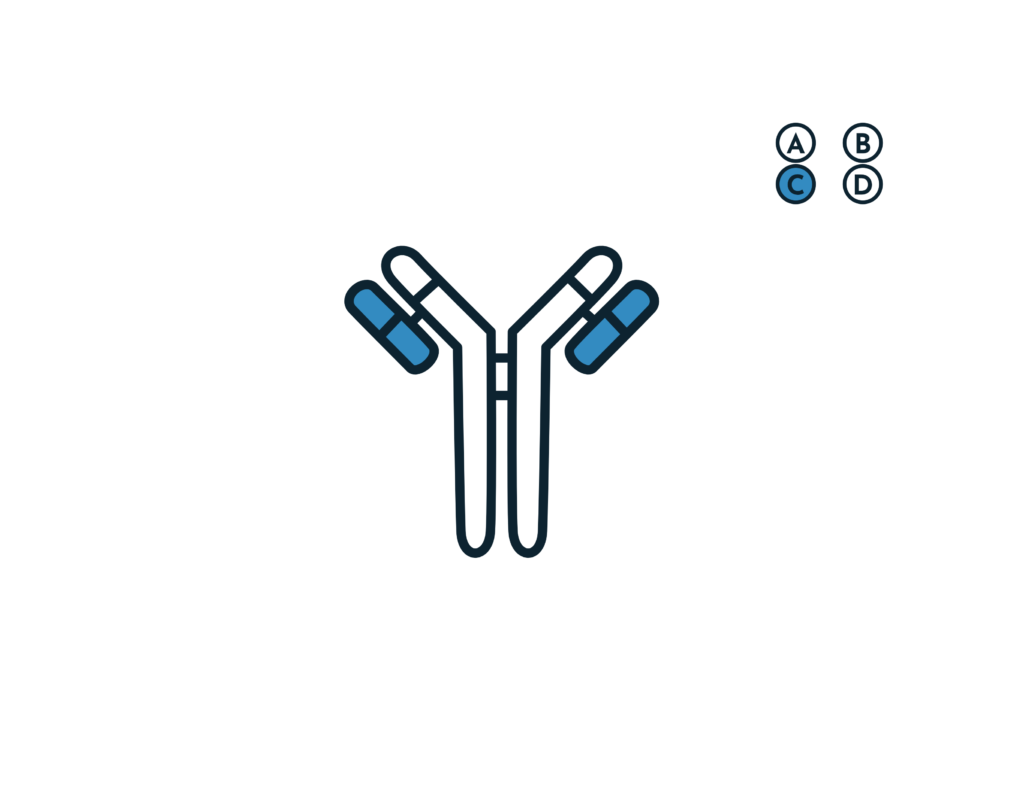
- Briefly describe Type III hypersensitivity
- Type III hypersensitivity is AKA “immune complex-mediated” hypersensitivity.
- Antibodies bind to antigens forming complexes, which circulate get stuck in vessels and stimulate inflammation.
- The end result is inappropriate inflammation and necrotizing vasculitis
- Diseases produced by immune complexes are those in which antigens persist without being eliminated as:
- Repeat exposure to extrinsic antigen
- Injection of large amounts of antigens
- Persistent infection
- Autoimmunity to self components
- List the diseases associated with Type III hypersensitivity reactions, their antigens and consequence (tissue affected)
- Systemic Lupus Erythematosus: Nuclear antigens → nephritis, skin lesions, arthritis
- Post-streptococcal glomerulonephritis: Streptococcal antigens → nephritis
- Polyarteritis Nodosa (PAN): Hepatitis B surface antigen → Systemic vasculitis
- Acute glomerulonephritis: Bacterial antigens (Treponema), Parasite antigens (Malaria, Schistosomes), Tumor antigens → Nephritis
- Reactive arthritis: Bacterial antigens (Yersinia) → Arthritis
- Serum sickness: Foreign proteins → arthritis, vasculitis, nephritis
- Arthus reaction: Foreign proteins; cutaneous vasculitis
- What are the types of Type III hypersensitivity reactions
- Systemic immune complex disease
- Complexes are formed in circulation and are deposited in several organs
- Includes:
- Serum sickness
- Meningitis
- Hepatitis
- Mononucleosis
- SLE
- RA
- Allergies to penicillins and sulfonamides
- Local immune complex disease
- Complexes are formed at sites of antigen injection and precipitate at the injection (local) site
- Includes: Arthus reaction
- Systemic immune complex disease
- Describe the pathophysiology of Type III hypersensitivity reactions
- Phases of Type III hypersensitivity
- Phase I: Immune complex formation
- Phase II: Immune complex deposition
- Phase III: Immune complex-mediated inflammation
- Activation of complement
- Neutrophil and monocyte chemoattraction
- Increased vascular permeability
- Anaphylatoxin – mediated activation of mast cells
- Complex-mediated phagocytosis and release of phagocyte granule enzymes and cytokines into the local microenvironment
- Fc receptor phagocytosis
- Release of proteases by neutrophils and monocytes
- PMN chemotaxis → Tissue damage
- Activate clotting
- Results in microthrombi – deposition, platelet aggregation
- Activation of complement
- Important complement fractions involved
- C3b: phagocytosis of complexes and bugs
- C3a, C5a (anaphylatoxins): increase permeability
- C5a: neutrophil and monocyte chemotaxis
- C5-9: Membrane damage and cytolysis (MAC)
- Phases of Type III hypersensitivity
- List the Hypersensitivity Pneumonitis Syndromes and their associated antigens
- Farmers lung: Thermophilic actinomycetes
- Malt worker’s lung: Aspergillus spores
- Pigeon fancier’s disease; Avian proteins
- Cheese washer’s lung: Penicillium spores
- Furrier’s lung: Fox fur
- Laboratory technician’s lung: Rat urine proteins
- Briefly describe Localized Type III reactions
- Injection of an antigen
- Leads to an acute Arthus reaction within 4-8 hours
- Localized tissue and vascular damage from edema, and erythema
- Severity varies from mild swelling to redness to tissue necrosis
- Insect bite
- May first have a rapid type I reaction
- 4-8 hours later a typical arthus reaction occurs
- Injection of an antigen
- Describe generalized Type III Hypersensitivity reactions
- Large amounts of antigen enter the bloodstream and bind to antibody → circulating immune complexes formed → cleared by phagocytosis or cause tissue damaging type III reactions
- What is the etiology of generalized Type III Hypersensitivity reactions
- Serum sickness → antigen is administered IV
- Infectious disease
- Meningitis
- Hepatitis
- Mononucleosis
- Drug reactions
- Allergies to penicillin and sulfonamides
- Autoimmune disease
- SLE
- RA
- Briefly describe the Arthus reaction
- Arthus reaction is a localized area of skin necrosis resulting from immune complex vasculitis.
- An antigen is injected into the skin of previously immunized persons causing pre-existing antibodies to form complexes with the antigens → The complexes precipitate at the site of infection and inflammation causes edema, hemorrhage and ulceration.
- Arthus reactions is a local immune complex deposition phenomenon eg. diabetic patients receiving insulin subcutaneously
- Local reactions: edema, erythema, necrosis
- Immune complex deposition in small blood vessels: vasculitis, microthrombi, vascular occlusion, necrosis
- Briefly describe Serum sickness
- Horse serum was used for immunization in the olden days.
- Injecting foreign antigens causes manufacture of antibodies, which form complexes with the antigens.
- The complexes lodge in the kidneys, joints, small vessels and the inflammation causes fever, joint pain and proteinuria.
- Serum sickness is a systemic immune complex phenomenon secondary to injection of large doses of foreign serum.
- Antigens are slowly cleared from circulation and immune complexes deposit at various sites.
- List drugs that are notorious for causing Serum sickness
- Cephalosporins, Ciprofloxacin, Furazolidone, Griseofulvin, Licomycin, Metronidazole, Para-aminosalicylic acids, penicillins, streptomycin, sulfonamides, tetracycline, allopurinol, barbiturates, bupropion, captopril, carbamazepine, fluoxetine, penicillamine
- What are the clinical features of Serum Sickness
- Purpuric rash, Erythema nodosum, Erythema multiforme
- 10 days post injection: fever, urticaria, arthralgia, lymphadenopathy, splenomegaly, glomerulonephritis
- What are the laboratory features of Serum Sickness
- CBC: Neutropenia, eosinophilia, thrombocytopenia
- Hepatitis serology: Asses Hepatitis B infection
- Inflammatory markers; Elevated ESR, CRP
- Complement levels: Low C3, C4 and CH50 levels
- Urinalysis: Mild proteinuria
- Skin lesions biopsy: Leukocytoclastic vasculitis that is non-specific
- What are the laboratory features of Hypersensitivity pneumonitis
- CBC: normal peripheral eosinophil
- IgG levels: Elevated
- Precipitating antibodies in serum against suspected causative agent
- CXR: Patchy or homogeneous bilateral interstitial and alveolar nodular infiltrates
- High-res CT: Fibrotic changes on CT
- Bronchoalveolar lavage: >50% lymphocytes
- What are the laboratory features of SLE
- ANA: sensitive
- Anti-dsDNA, anti-smith: Specific
- Urinalysis with microscopy and urine spot protein to creatinine ratio: evaluate possible nephritis and quantification of proteinuria
- Renal biopsy: definitive diagnosis and classification of lupus nephritis
- Appropriate imaging: Evaluate pulmonary and joint symptoms


