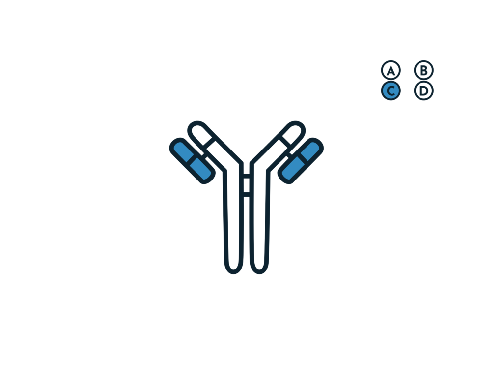
- Describe the mechanism of Type II hypersensitivity
- Opsonization and phagocytosis
- Opsonized RBCs and platelets become targets for phagocytosis by neutrophils and macrophages.
- Phagocytes express Fc receptors for IgG and Breakdown products of C3
- Opsonized cells are eliminated in the spleen, and this is why splenectomy is beneficial in autoimmune thrombocytopenia and some forms of autoimmune hemolytic anemia
- Inflammation
- Antibodies bound to cellular or tissue antigens activate the complement cascade via the classical pathway
- Products of complement activation recruit neutrophils and monocytes triggering inflammation in tissues.
- Severe damage to membranes is caused by protease released from leukocytes attracted to the site by C5a
- Antibody mediated cellular dysfunction
- Antibodies directed against cell surface receptors impair or dysregulate cellular function without causing cell injury or inflammation
- Antibodies can also stimulate excess cellular responses (Graves disease)
- Antibodies against hormones and other essential proteins can neutralize and block the actions of these molecules causing functional derangement
- Opsonization and phagocytosis
- List Type II hypersensitivity reactions
- Hemolytic disease of the newborn
- immune mediated hemolytic anemia
- Idiopathic thrombocytopenic purpura
- Acute hemolytic transfusion reaction
- Goodpasture syndrome
- Graves disease
- Myasthenia gravis
- Briefly describe Immune Hemolytic anemia
- Antibodies against RBCs may be autoantibodies or alloantibodies, or antibodies directed against a drug or its metabolite.
- They lead to RBC destruction via complement mediated lysis, opsonisation, or both mechanisms.
- The main antibody isotypes involved include IgG and IgM
- IgM are highly effective at activating complements (unlike IgG which require a sufficient number of moleculess on RBC surface).
- MAC formation lyses RBCs while C3b facilitates opsonisation and destruction by the RES (mainly by kupffer histiocytes in the liver)
- IgG antibodies mediate hemolysis predominantly by extravascular mechanisms, causing opsonized RBCs to be eliminated by splenic macrophages
- Spherocytes are the result of IgG mediated phagocytosis as part of the membrane is removed.
- Spherocytes are eventually removed from circulation via entrapment in the cords of billroth, where they are rapidly phagocytized by macrophages.
- What are the laboratory features of Immune Hemolytic Anemia
- CBC – Decreased hemglobin
- Elevated Reticulocyte count
- PBF – Polychromasia, Spherocytes, RBC agglutination in IgM mediated hemolysis
- Increased serum indirect bilirubin
- Elevated LDH
- Positive DAT
- Briefly describe Autoimmune Hemolysis
- Autoimmune HA is characterized by a positive Coombs (DAT) which detects antibodies, with or without complement on the RBC surface.
- Warm agglutinin(IgG): Bind to RBCs more strongly at 37*C and have decreased affinity at lower temperatures
- Cold agglutinin (IgM): Autoantibodies bind to red cells more strongly at 4*C, with little affinity at physiologic temperatures
- Warm and cold autoantibodies may coexist.
- Briefly describe Warm Autoimmune Hemolytic Anemia (AIHA)
- Affects all age groups.
- F>M
- .Caused by polyclonal IgG molecules produced by polyclonal B cells.
- Rarely caused by IgA and IgM.
- Hemolysis is predominantly extravascular.
- Clinical presentation depends on the speed of development of anemia, capacity of bone marrow compensation and the effects of the underlying disease
- What is the etiology of Warm AIHA
- Primary: Idiopathic
- Secondary: Systemic autoimmune diseases (RA, SLE), Lymphoproliferative neoplasms (CLL, Lymphoma, Thymoma), Infections (EBV), Tumors (Ovarian carcinoma)
- What are the clinical features of Warm AIHA
- Symptoms of anemia: Fatigue, dizziness, dyspnea; Fever, Jaundice, Splenomegaly, Hepatomegaly
- What are the laboratory features of Warm AIHA
- CBC: Low Hb, Normal or elevated MCV, Increased RDW, Increased WBC or platelets
- Elevated Retics
- PBF: Polychromasia, Spherocytes, nRBCs, Red cell agglutination
- Increased indirect Bili
- Reduced haptoglobin
- Urine: Hemoglobinuria
- Increased LDH
- Positive DAT against IgG/ C3d
- How is warm AIHA treated
- Supportive: Blood transfusion
- Corticosteroids
- Splenectomy: if response to steroids fails
- Monoclonal antibodies
- Cytotoxic immunosuppressive drugs: Azathioprine (antimetabolites)
- Briefly describe cold AIHA
- Caused by cold agglutinins (IgM) that react optimally at 4*C, usually monoclonal and occur at high titers.
- These antibodies are also capable of reacting at temperatures greater than 30C and bind to RBC antigens near or at 37C.
- Cold agglutinins bind to RBCs particularly in the peripheral circulation and vessels of the skin where temperatures can drop to 30*C.
- IgM autoantibodies activate the classical complement pathway.
- Hemolysis is predominantly extravascular by Hepatic Kupffer histiocytes via C3b, however at high thermal amplitude there is full deficiency in complement regulators, full complement activation and activation of intravascular hemolysis
- What is the etiology of cold AIHA
- Primary: Idiopathic
- Secondary: Infections (Mycoplasma pneumoniae), Lymphoproliferative disorders (Multiple Myeloma, CLL/SLL etc.)
- Briefly describe Alloimmune Hemolytic Anemia
- Alloimmune HA results from alloantibodies
- Prototypes
- Hemolytic transfusion reactions
- Hemolytic disease of the newborn
- Briefly describe Hemolytic Transfusion Reactions
- A complication of blood transfusion, there is mediated destruction of donor cells via IgM or IgG.
- Hemolysis is either intravascular or extravascular, and can be acute or delayed
- Briefly describe Acute hemolytic transfusion reaction (AHTR)
- Acute hemolytic transfusion reactions occur up to 1-2 hours post transfusion.
- Common cause is a clerical error and is related to ABO incompatibility resulting in the rapid destruction of the donor RBCs by host antibodies.
- Most severe reactions involve group A RBCs given to a patient who is group A
- Describe the pathophysiology of AHTR
- Intravascular hemolysis occurs due to ABO compatibility
- ABO antibodies fix complement leading to MAC formation and rapid lysis
- Other antibodies (especially Kidd) may also fix complement and lyse RBCs
- Hemolysis leads to
- Release of free HFB and HGB free RBC stroma into circulation
- Stimulation of intrinsic coagulation pathway and bradykinin via Ag-Ab complexes
- C3a and C5a generation
- Production of several important cytokines – TNF-a, IL-1B,IL-8, IL-6
- TNF-a , IL-1B, and IL-6 strongly promote fever and activate and stimulate WBCs
- Direct intrinsic pathway activation by Ag-Ab complexes interact with factor X12
- Indirect activation of the extrinsic pathway by TNF-a which stimulates Tissue factor
- Decreased Protein C inhibition via decreased thrombomodulin
- Tissue factor activation predisposes to DIC in 10% of patients
- Anaphylatoxins promote bronchoconstriction from histamine release (Wheezing/ dyspnea)
- Sympathetic response to hypotension leads to renal vasoconstriction, free Hb scavenges renal NO promoting vasoconstriction, Renal microthrombi from DIC also decrease renal blood flow contributing to the risk of Acute Tubular Necrosis
- What are the clinical features of AHTR
- Fever and chills
- Infusion site pain
- Hypotension or shock
- Hemoglobinuria (first indication of hemolysis in anesthetized patients)
- DIC or increased bleeding (important in anesthetized patients),
- Sense of “impending doom”
- What are the Laboratory features of AHTR
- Hemoglobinemia: pink or red serum or plasma; lasts several hours in those with adequate renal function
- Hemoglobinuria
- Positive Direct Antiglobulin Test
- Elevated indirect and direct bilirubin
- Lab features of DIC (D-dimers, Decreased fibrinogen, etc.)
- PBF: Schistocytes (intravascular hemolysis), Spherocytes (IgM mediated extravascular hemolysis)
- Low haptoglobin
- Briefly describe Delayed Hemolytic Tranfusion reaction (DHTR)
- DHRT occurs days to weeks post-transfusion in patients who have been alloimmunized by blood transfusion or prior pregnancy.
- On second exposure an anamnestic response is mounted against duff/kidd antigens by IgG antibodies (Reactive at 37 C).
- Hemolysis is extravascular, with or without complement fixation
- Lab features
- Positive DAT
- Rise in unconjugated hemoglobin
- Failure of Hb to rise post-transfusion
- Briefly describe Hemolytic Disease of the Newborn
- IgG alloantibodies produced by the mother cross the placenta and bind to fetal RBCs containing Rh, ABO, Kell and other minor antigens.
- Sensitized RBCs are cleared by splenic macrophages and hemolysis develops.
- There is erythroid hyperplasia in the fetal marrow and extramedullary erythropoiesis in the fetal spleen, liver, kidneys and adrenals.
- nRBC are released into the circulation (Hence erythroblastosis fetalis).
- Severe anemia in utero leads to generalized edema, ascites and hydrops fetalis
- Lab features in a newborn
- CBC: decreased Hb
- Elevated reticulocytes
- Elevated serum indirect Bili
- PBF: Polychromasia, nRBCs
- Positive DAT
- Briefly describe Drug Induced Hemolytic Anemia
- Drugs have a molecular weight that is too low to make them immunogenic (haptens) unless they are conjugated with large carrier molecules (proteins) that allow them to elicit and immune response
- Pathogenesis
- Drug adsorption and extravascular hemolysis (IgG): Penicillins and cephalosporins
- Immune complex and complement activated intravascular hemolysis (IgG/ IgM): Rifiampicin, Quinin, Hydrochlorothiazide – IgM/IgG)
- RBC autoantibody induction: Drug induces the patient to produce warm agglutinin IgG autoantibodies against RBC self antigens → Extravascular hemolysis
- Membrane modification (Cephalosporins): Modify red cell membrane proteins causing IgG and complement to adsorb on the cell surface → non-immune drug induced hemolysis
- Treatment: Discontinue drug, Blood transfusion if anemia is severe, Plasma exchange
- Briefly describe Immune Thrombocytopenia (formerly Idiopathic Thrombocytopenic Purpura; ITP)
- The term idiopathic no longer applies (caused by immune dysregulation) and purpura has been abandoned as it is misleading (1/3 of patients diagnosed with ITP have no bleeding despite low platelet counts)
- It is a syndrome in which platelets become coated with autoantibodies to platelet membrane antigens, resulting in splenic sequestration and phagocytosis by mononuclear macrophages
- Etiology and epidemiology
- Can be acute or chronic
- Acute ITP is primarily a disorder of children and usually follows a non-specific URTI or GIT infection. Children produce antibodies and immune complexes against viral antigens and platelet destruction results from the binding of these antibodies or immune complexes to the platelet surface.
- The chronic form affects individuals between ages 20-50 with a female preponderance. It is usually no preceded by a viral infection
- Describe the pathophysiology of Immune Thrombocytopenia
- An abnormal autoantibody usually IgG with specificity for one or more platelet membrane glycoproteins (Gp IIb, IIIa, Gp Ib/IX/V) binds to circulating platelet membranes
- The antibodies are formed in the white pulp of the spleen
- Autoantibody-coated platelets induced Fc receptor-mediated phagocytosis by mononuclear phagocytes in the spleen and liver
- What are the clinical features of Immune Thrombocytopenia
- Purpura, petechiae, ecchymoses
- Oozing from venu-puncture sites
- Gingival bleeding
- Features of intracranial hemorrhage: headache, blurred vision, somnolence, loss of consciousness
- Signs of chronic disease, infection, wasting, or poor nutrition indicated thrombocytopenia due to other causes. Splenomegaly excludes the diagnosis of ITP.
- What are the laboratory features of Immune Thrombocytopenia
- CBC: Isolated thrombocytopenia (Hallmark of ITP)
- PBF: Normal platelet morphology, varying numbers of large platelets, esp in acute cases (BM compensation), Normal RBC and leukocytes
- Bone Marrow aspirate/ Biopsy: Cellularity and morphology of erythroid and myeloid precursors should be normal, the number of megakaryocytes may be increased
- Assay for anti-platelet antibodies is positive
- Briefly describe Grave’s disease
- Graves disease is an autoimmune disease that primarily affects the thyroid gland.
- It may also affect other organs including the eyes and skin.
- It is the most common cause of hyperthyroidism (60-80%) and is common in 20-50y.
- Graves is F>M.
- In some patients, Graves disease represents a part of more extensive autoimmune processes leading to dysfunction of multiple organs (Polyglandular autoimmune syndromes)
- What other diseases is Grave’s disease commonly associated with
- Pernicious anemia
- Vitiligo
- Type I Diabetes Mellitus
- Autoimmune adrenal insufficiency
- Systemic sclerosis
- Myasthenia gravis
- Sjorgen syndrome
- Rheumatoid arthritis
- Systemic lupus erythematosus (SLE)
- What is the etiology of Grave’s disease
- Genetic susceptibility: HLA-DRB1, HLA-DQB1, CD40, CTLA4, PTPN22
- Environmental triggers: Infection, stress, smoking, iodine exposure, drugs
- Describe the pathogenesis of Grave’s disease
- Graves B and T lymphocyte mediated autoimmunity directed at Thyroglobulin, Thyroid peroxidase, Sodium Iodide symporter and Thyrotropin receptor (MAIN)
- Thyrotropin is the primary autoantigen and is responsible for the manifestation of hyperthyroidism
- IgG1 is implicated
- Thyroid gland is under constant stimulation by autoantibodies
- Antibodies stimulate both Thyroxine synthesis and thyroid gland growth, causing hyperthyroidism and thyroidomegaly
- The anti-sodium-iodide symporter, antithyroglobulin, and anti-thyroid peroxidase antibodies appear to have little role in the etiology of hyperthyroidism in Graves, However they are markers of autoimmune disease against the thyroid gland
- Intrathyroidal lymphocytic infiltration is the initial histologic abnormality in persons with autoimmune thyroid disease and can be correlated with the titre of thyroid antibodies
- What are the clinical features of Grave’s disease
- Graves orbitopathy (ophthalmopathy) caused by inflammation, cellular proliferations, increased growth of extraocular muscles and retro-orbital CT and adipose tissue due to the actions of thyroid stimulating antibodies and cytokines release by T lymphocytes (Killer cells)
- These cytokines and thyroid stimulating antibodies activate
- Proptosis
- General – fatigue, general weakness
- Dermatological: warm, moist, fine skin; seating; fine hair onycholysis; vitiligo alopecia; pretibial myxedema
- Neuromuscular: tremors, proximal muscle weakness, easy fatigability, periodic paralysis in persons of susceptible ethnic groups
- Skeletal: Back pain, increased risk of fractures
- Cardiovascular: palpitations, dyspnea on exertion, chest pain, edema
- Respiratory: dyspnea
- Gastrointestinal: increased bowel motility with increased frequency of BMS
- Ophthalmology: tearing, gritty sensation in the eye, photophobia, eye pain, protruding eye, diplopia, visual loss
- Renal: polyuria, polydipsia
- Hematologic: easy bruising
- Metabolism: heat intolerance, weight loss despite increase or similar appetite, worsening diabetes control
- Endocrine or reproductive: irregular menstrual periods, decreased menstrual volume, secondary amenorrhea, gynecomastia, impotence
- Psychiatric: restlessness, anxiety, irritability, insomnia
- What are the laboratory features of Grave’s disease
- TFT: Low TSH, Elevated T3/T4 (Subclinical hyperthyroidism – TSH low, T3/T4 normal)
- Measurement of TSH receptor antibody (TRAb)
- Measurement of other antibodies: Antithyroglobulin antibodies, antithyroidal peroxidase antibodies, anti-sodium-iodide symporter antibody
- Radioactive iodine uptake scan (RAIU) using I-123 or I-131: Uptake is high and diffuse in grave’s disease
- Histology: thyroid glands show lymphocytic infiltrates and follicular hypertrophy, with little colloid present
- Briefly describe Goodpasture Syndrome
- Goodpasture syndrome is caused by autoantibodies directed against collagen in the basement membrane of the kidneys and lungs. Etiology is unknown, but thought to follow a viral infection.
- Risk factors
- HLA-DR2 (Genetic susceptibility)
- Pathogenesis
- Cytotoxic antibodies activate complement
- C5a causes neutrophil chemotaxis
- Neutrophils release enzymes that damage te kidney and lung tissue
- Clinical findings
- Hematuria
- Proteinuria
- Pulmonary hemorrhage
- Diagnosis
- Detection of antibody and complement bound to basement membranes in fluorescent antibody test (microscopy)
- Briefly describe Myasthenia gravis
- Myasthenia gravis is a relatively acquired autoimmune disorder caused by antibody mediated blockade of neuromuscular transmission resulting in skeletal muscle weakness.
- Autoantibodies are formed against the nACh receptors at the NMJ
- This prevents nerve impulses from triggering muscle contractions. Commonly affects the muscles of the eyes, face, and swallowing
- Clinical presentation
- Proggresive weakness
- Ptosis
- Diplopia
- Dysphagia
- Paralysis of respiratory muscles


