Last updated:
November 12, 2024
- What is the fluid mosaic model of the plasma membrane
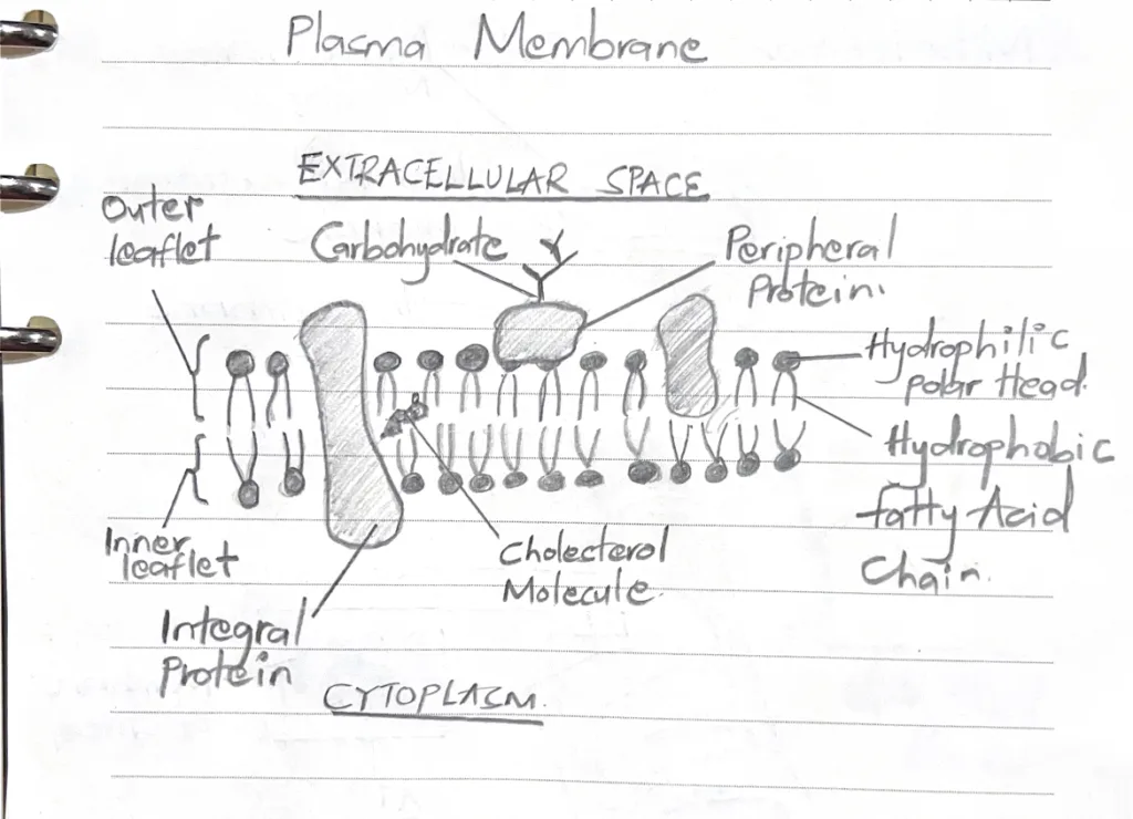
- The plasma membrane is a lipid bilayer primarily consisting of phospholipids, cholesterol and proteins.
- Hydrophobic fatty acid chains form the inner layer while hydrophilic phosphate groups form the outer layers (the extracellular and intracellular surfaces)
- Cholesterol is incorporated within the gaps between phospholipids equally on both sides
- Lipid rafts (cholesterol and glycosphingolipid rich areas) exist
As regards membranous proteins, distinguish between carrier proteins, channel proteins, and pumps
| Carrier proteins | Channels | Pumps | |
|---|---|---|---|
| Function | Binds specific molecules and changes their conformation to transport them across the cell membrane | Allows passive diffusion of ions, small molecules and water across the cell membrane | Actively transports ion such as Na+ across the cell membrane |
| Form of transport | Passive transport | Passive transport | Active transport |
- Describe the cell cycle
- G1 phase: Begins at the end of the M phase. It is the longest phase. The cell gathers nutrients and synthesizes RNA and proteins necessary for DNA and chromosome replication
- S phase: DNA is replicated
- G2 phase: The cell prepares for division by checking replicated DNA
- M phase: Mitosis occurs in this phase. Includes both karyokinesis and cytokinesis. The M phase is divided into Prophase, Metaphase, Anaphase, and Telophase
- Describe modes of bulk transport across the plasma membrane
- Endocytosis: Vesicular transport in which substances enter the cell
- Pinocytosis: Non-specific uptake of fluids and small proteins through vesicles. Clathrin-independent endocytosis.
- Phagocytosis: Ingestion of large particles (cell debris, bacteria). Cells send out pseudopodia and engulf the material in vacuoles known as phagosomes.
- Receptor-mediated endocytosis: Specific uptake of molecules into the cells upon recognition by “cargo receptors”. Clathrin-dependent endocytosis.
- Exocytosis: Vesicular transport in which substances leave the cell
- Constitutive pathway: Substances designated for exocytosis are continuously delivered to the cell membrane. Cells lack secretory granules.
- Regulatory secretory pathway: A hormonal or neural stimulus is required to activate exocytosis of substances.
- Endocytosis: Vesicular transport in which substances enter the cell
- What is the structural make-up of the microtubule? What is an MTOC?
- Microtubule structure: Microtubules are elongated polymers composed of dimeric tubulin molecules (equal parts a-tubulin and B-tubulin). Form a 9 + 2 arrangement in cilia whereby 9 microtubules form a circle surrounding 2 central microtubules.
- Microtubule Organizing Centre (MTOC or Centrosome): Regions in the cell that contain centrioles and pericentriolar material. Most microtubules are formed at the MTOC and are directed to different cellular areas. Contain y-tubulin rings that form the nucleation point for the assembly of a-tubulin and B-tubulin molecules
- Name 5 lysosomal storage diseases
- Niemann Pick Disease
- Gaucher Disease
- Fabry Disease
- Krabbe disease
- Tay-Sachs disease
- Give examples of inclusions in cells
- Lipofuscin: Brownish-yellow “wear-and-tear” pigment
- Hemosiderin: Iron storage complex. Deep brown granules in light microscopy (Indistinguishable from Lipofuscin. Must stain with Perl’s Prussian Blue)
- Glycogen: Branched polymer of glucose molecules. Special staining by PAS or Toluidine blue.
- Lipid inclusions (fat droplets): Appear as “holes” in the cytoplasms, representing sites where lipids were removed during tissue preparation.
- Crystalline inclusions: contained in specific cells, as recognized in light microscopy.
- List 5 features of active protein synthesizing cells
- rER tends to be profuse and to form closely packed parallel laminae of flattened cisternae.
- Nucleus typically contains a prominent nucleolus
- Chromatin in the nucleus is mainly dispersed (euchromatin)
- Numerous ribosomes stud the surface of the rER membrane
- Many ribosomes lying free in the intervening cytosol
- Basophilic clumps in the cytoplasm represent areas of plentiful rER.
- State the structural features of cells undergoing apoptosis
- DNA fragmentation
- Decrease in cell Volume
- Loss of Mitochondrial function
- Membrane blebbing
- Formation of Apoptotic bodies
- Name the organelle associated with the following diseases: Cystic fibrosis, Myopathies, Alzheimer’s disease, Zellweger’s syndrome, Polycystic Kidney Disease
- Cystic fibrosis: Plasma membrane
- Myopathies: Proteasomes, Glycogen
- Alzheimer’s disease: Cytoskeleton, Ribosomes, Proteasomes
- Zellweger’s syndrome: Peroxisome
- Polycystic Kidney Disease: Golgi apparatus
- With the aid of a diagram, describe the structure of the cell membrane
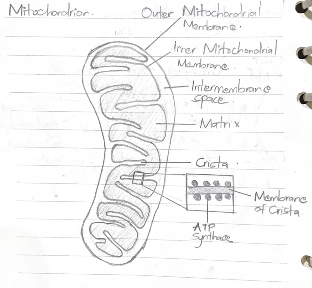
- With the aid of a labelled diagram, describe the structure of mitochondria
- Describe the classification of somatic cells with reference to cell renewal
- Rapidly renewing cell populations: Name suggests. Include blood cells, Epithelial cells, Dermal fibroblasts of the skin and epithelial cells and subepithelial fibroblasts of the GI mucosa
- Slowly renewing cell populations: These cells slowly increase in size during life – Smooth muscle cells of most hollow organs (GI tract), Fibroblasts of the uterine wall, epithelial cells of the lens of the eye
- Renewing cell populations: Slow or rapidly renewing cells that display regular mitotic activity. Division results in 2 daughter cells that differentiate morphologically and functionally or 2 stem cells. Daughter cells eventually reach their mitotic limit.
- Stable cell populations: Cells that divide episodically and slowly to maintain normal tissue or organ structure. They may be stimulated by injury to divide. Periosteal cells, Perichondrial cells, Smooth muscles cells, Endothelial cells, Fibroblasts
- Static cell populations: Cells that no longer divide (post-mitotic cells) – Neurons, Skeletal Muscles, Cardiac Muscles
- State two clinical correlates related to cell renewal
- Hepatocytes regenerating after liver transplant (Stable cell population)
- Wound healing through regeneration of skin epithelial cells and dermal fibroblasts (Rapidly renewing cell populations)
- Describe the different pathways of autophagy
- Macroautophagy (Autophagy): A non-specific process. An entire organelle or portion of the cytoplasm is surrounded by a double membrane of endoplasmic reticulum (the isolation membrane) to form a vacuole known as the autophagosome. Lysosomal enzymes are delivered to the autophagosome converting it into a lysosome. Hydrolytic enzymes degrade the isolation membrane and the organelle. Macroautophagy occurs in the liver during the first stages of starvation.
- Microautophagy: Also a non-specific process. Cytoplasmic proteins are degraded in a slow continuous process. These small soluble proteins are internalized into lysosomes through invagination of the lysosomal membrane.
- Chaperone-mediated autophagy: A specific or selective process. It is activated during nutrient deprivation and requires the presence of targeting signals on the proteins to be degraded and a specific receptor on the lysosomal membrane. Heat-shock proteins bind to the protein and assist in their transportation across the lysosomal membrane. Chaperone-mediated autophagy is responsable for the degradation of approximately 30% of cytoplasmic proteins in the liver and kidneys.
- State one clinical correlate related to autophagy
- The lysosomal storage disorder Inclusion-cell disease (I-cell disease) – Lysosomal hydrolyses are not present in lysosomes causing impaired degradation and the accumulation of abnormal lysosomal contents in serum
- Briefly describe the functions of the following organelles: Mitochondria, Lysosomes, Peroxisomes, Ribosomes, Nucleolus, Rough Endoplasmic Reticulum, Smooth Endoplasmic Reticulum, Golgi apparatus
- Mitochondria ATP production, calcium homeostasis, apoptosis
- Lysosomes Intracellular digestion/ breakdown of macromolecules
- Peroxisomes Oxidation and detoxification
- Ribosomes Translation of mRNA (protein synthesis)
- Nucleolus Synthesis of ribosomes
- Rough endoplasmic reticulum Synthesis of proteins (translation and post translational modification)
- Smooth endoplasmic reticulum Synthesis of Lipids and steorids, detoxification
- Golgi apparatus Sorting, packaging and transport of materials in vesicles
- What are the major functions of the following non-membranous organelles: Actin filaments, Intermediate filaments, Microtubules
- Actin filaments
- Anchoring and moving membrane proteins
- Form the structural core of microvili
- Locomotion of cells
- Extension of cell processes
- Intermediate filaments
- Formation of cell-to-cell junction
- Formation of the cell-to-extracellular matrix junction
- Microtubules
- Movement of intracellular organelles via molecular motor proteins (Dynein and Kinesisn) associated with microtubules
- Actin filaments