Retinoblastoma is a neuroectodermal malignancy arising from retinal cells. It is one of the small round blue cell tumors of childhood. It is caused by a mutation of the retinoblastoma gene (RB1) located on chromosome 13q14. Children born with germline mutations of RB1 are also at risk of other primary malignancies (e.g. Osteosarcoma). The best initial step in diagnosing retinoblastoma is an immediate ophthalmoscopic examination under general anaesthesia. Biopsy of the tumor is not performed due to the risk of seeding the vitreous.
Type
Pathogenesis
Presentation
Heritable Retinoblastoma
The child is born with one germline mutation of the RB1 gene. The other is acquired after birth resulting in a tumor (Two-hit hypothesis).
Presents earlier and tends to be bilateral.
Non-heritable Retinoblastoma
The child is born with no RB1 mutation and acquires somatic mutations of both RB1 genes after birth
Presents relatively later and tends to be unilateral.
Accounts for about 2.5% of all childhood cancers from birth to 14 years. Affects 1 in 15,000 children. Median age of diagnosis is 15 months of age. 90% of cases are diagnosed before 5 years of age. Overall, 70-80% are unilateral. 20-30% are bilateral. Of heritable retinoblastoma, 90% of inherited mutations develop spontaneously. 10% of inherited mutations come from the parent.
What are the other small round blue cell tumors of childhood?
Hyphema (due to destruction of the anterior chamber)
Investigations
Ophthalmoscopic examination under general anesthesia
The tumor appears as a whitish-pink creamy mass
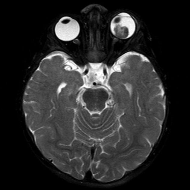
CT-scan or MRI of the orbit: to determine extra-ocular extension, infiltration, and involvement of the optic nerve
Bilateral Bone Marrow Biopsy: for prognosis after diagnosis
CSF cytology: for prognosis after diagnosis
Treatment
External beam radiation
Neoadjuvant chemotherapy can be used to reduce tumor bulk before radiation (using Carboplatin, Vincristine, and Etoposide)
Chemotherapy for metastatic disease
Enucleation if radiation and chemotherapy fail or if there is no vision in the affected eye (non-salvageable)
5-years survival
90% if confined to the orbit
40% if invades the optic nerve (CN II)
Rare if there are distant metastasis
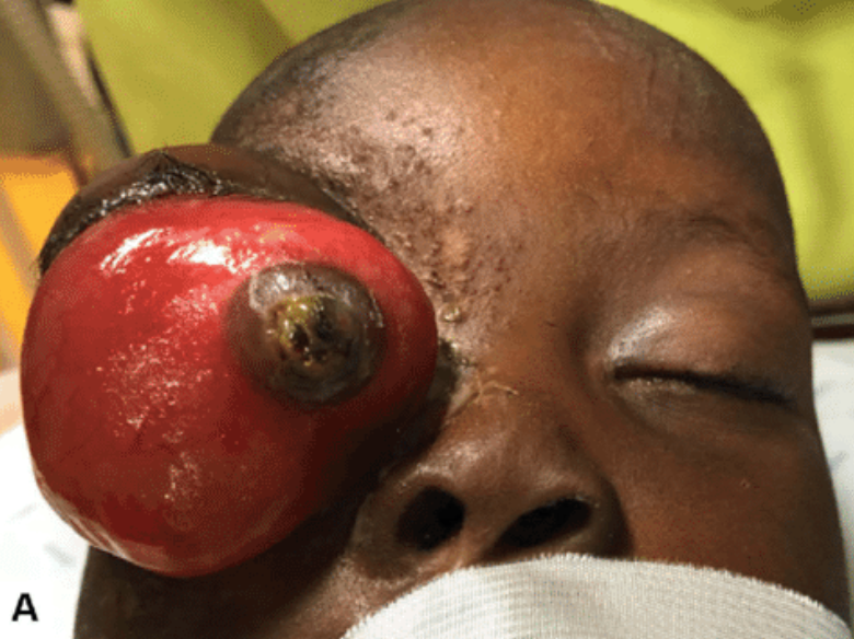
Leukocoria seen in the right eyeLeukocoria with tumor visible through the right pupil. Source:New York Eye Cancer CenterHyphema can be seen if the tumor invades the anterior chamber. Source: Tim Root Virtual Eye ProfessorOrbital invasion of the right eye. Source- Treatment of advanced retinoblastoma in Children evacuated from Low-Income CountriesRetinoblastoma on FundoscopyRetinoblastoma left-eye seen on T2-weighted MRI. Source: Radiopaedia
To provide the best experiences, we use technologies like cookies to store and/or access device information. Consenting to these technologies will allow us to process data such as browsing behavior or unique IDs on this site. Not consenting or withdrawing consent, may adversely affect certain features and functions.
Functional
Always active
The technical storage or access is strictly necessary for the legitimate purpose of enabling the use of a specific service explicitly requested by the subscriber or user, or for the sole purpose of carrying out the transmission of a communication over an electronic communications network.
Preferences
The technical storage or access is necessary for the legitimate purpose of storing preferences that are not requested by the subscriber or user.
Statistics
The technical storage or access that is used exclusively for statistical purposes.The technical storage or access that is used exclusively for anonymous statistical purposes. Without a subpoena, voluntary compliance on the part of your Internet Service Provider, or additional records from a third party, information stored or retrieved for this purpose alone cannot usually be used to identify you.
Marketing
The technical storage or access is required to create user profiles to send advertising, or to track the user on a website or across several websites for similar marketing purposes.