The vagina is a cylindrical tube that runs from the cervix to the vulva. Rugae provide a stretching probability which is useful during childbirth.
#1 cause of dyspareunia in a post-menopausal woman
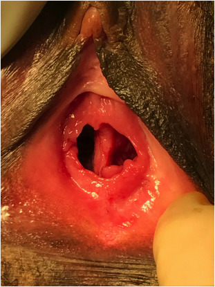
Post-menopausal atrophic vaginitis
Vaginal Foreign Body
Something in the vagina that is not supposed to be there. Commonly seen in children. The most common culprits are toilet tissue, tampons and sexual devices. Finding a vaginal foreign body warrants a pelvic exam. Sexual abuse should be ruled out.
Signs and symptoms
Vaginal bleeding
Foul-smelling vaginal discharge
Differentials for paediatric vaginal bleeding
Urethral prolapse
Sexual abuse
Foreign body
Accidental trauma
Epidermal sinus tumors
Rhabdomyosarcoma
Hemangioma
Management
Pelvic exam and rectal exam (with gentle anterior pressure to expel foreign bodies)
Speculum to inspect for mucosal damage
Liberal irrigation
Large or sharp objects may require general anaesthesia
Paediatric vaginal foreign bodyVaginal foreign body
Gartner’s Duct Cyst
It is formed when the wollfian duct does not fully involute. Gartner’s duct cyst is the most common benign cystic lesion of the vagina. Usually asymptomatic and is discovered incidentally on transvaginal ultrasound. Located on the anterolateral wall of the proximal vagina.
Gartner’s duct cyst
Signs and Symptoms
Dyspareunia
May cause obstetric complications
May be associated with Genitourinary anomalies
Other cystic anomalies in the female genital tract
Cystic abnormality
Description
Bartholin’s gland cyst
Asymptomatic or Pelvic pressure. Visualized at 4 or 8 o clock position
Vaginal inclusion cyst
May occur anywhere in the vagina following trauma
Skene’s duct (paraurethral) cyst
Asymptomatic, rarely urinary outflow obstruction
Nabothian cyst
Asymptomatic, raised white-yellow lesions picked up on cervical exam
Atrophic Vaginitis
Thinning of the vaginal epithelium due to decreased estrogen levels. Very common in post-menopausal women. Can occur in those who are functionally post-menopausal (following BSO) or women on anti-estrogens (Endometriosis – Danazol or Leuprolide, Breast cancer – Tamoxifen, Progestin-only contraceptives). Rule out exogenous agents (soap, perfume e.t.c). Consider vulvovaginitis if there is discharge.
Transverse vaginal septum: Due to incomplete fusion of the mullerian duct and urogenital sinus. Asymptomatic and noticed on physical exam. Presents as primary amenorrhea (”cryptomenorrhea”) due to uterine outflow obstruction – cyclical lower abdominal pain, amenorrhea, central pelvic mass (hematometra or hematocolpos). Incomplete septa may have oligomenorrhea, dyspareunia or obstetric complications.
Longitudinal vaginal septum: Due to incomplete fusion of the mullerian . Asymptomatic and noticed on physical exam. May be associated with uterine septum or uterine didelphys which causes obstetric complications (miscarriage, abnormal implantation e.t.c due to associated mullerian duct abnormalities)
To provide the best experiences, we use technologies like cookies to store and/or access device information. Consenting to these technologies will allow us to process data such as browsing behavior or unique IDs on this site. Not consenting or withdrawing consent, may adversely affect certain features and functions.
Functional
Always active
The technical storage or access is strictly necessary for the legitimate purpose of enabling the use of a specific service explicitly requested by the subscriber or user, or for the sole purpose of carrying out the transmission of a communication over an electronic communications network.
Preferences
The technical storage or access is necessary for the legitimate purpose of storing preferences that are not requested by the subscriber or user.
Statistics
The technical storage or access that is used exclusively for statistical purposes.The technical storage or access that is used exclusively for anonymous statistical purposes. Without a subpoena, voluntary compliance on the part of your Internet Service Provider, or additional records from a third party, information stored or retrieved for this purpose alone cannot usually be used to identify you.
Marketing
The technical storage or access is required to create user profiles to send advertising, or to track the user on a website or across several websites for similar marketing purposes.