Overview
Approximately 25% of pregnancies experience bleeding in the first trimester. Most women need to be reassured since 50% of the time the pregnancy will go on uncomplicated. Lower abdominal pain and heavy bleeding are associated with an increased risk of pregnancy loss. The vast majority of losses are due to chromosomal anomalies. Every patient presenting with early pregnancy bleeding should get their blood grouped (and possibly crossmatched), and a sterile speculum exam.
Important first steps in the management of first-trimester bleeding (or bleeding after secondary amenorrhoea) include IV fluids, physical exam + sterile speculum exam, quantitative B-hCG, Complete Blood Count, and transvaginal ultrasound. hCG normally takes 2-4 weeks to clear after evacuation.
Progesterone level of <6 ng per mL (19.1 nmol per L) reliably excludes a viable pregnancy (with a Negative Predictive Value of 99%)
- Differentials for early pregnancy bleeding
- Ectopic pregnancy
- Threatened miscarriage (50% will go on to have a normal pregnancy)
- Miscarriage
- Inevitable miscarriage
- Incomplete miscarriage
- Complete miscarriage
- Missed miscarriage
- Molar pregnancy
- Gynaecological causes:
- Vulvar, vagina, or cervical trauma
- Cervical polyp
- Cervical ectopia
- Cervical cancer
- Trichomoniasis
- Candidiasis
- Venereal warts
- What are the possible causes of pallor in a patient with early pregnancy bleeding?
- Vasovagal shock
- Hypovolemic shock
- Anemia
- Septic shock
General Management of Early Pregnancy Bleeding
- Physical examination
- Bimanual exam: to feel for masses e.g. adnexal mass, cervical motion tenderness,
- Sterile Speculum exam
- Check whether the cervical os is dilated or closed
- Look for clots or products of conception
- Rule out vulvar, vaginal, or cervical trauma
- Investigations
- Quantitative B-hCG: compare past and future measurements. Should double every 48 hours. Compared to estimated dates on LMP
- A normal pregnancy with serum hCG > 1,500IU/L should be able to be confirmed as intrauterine on transvaginal ultrasound
- CBC: to evaluate blood loss
- Transvaginal ultrasound: to visualize the fetus
- Gestational sac visible at 5 weeks
- Yolk sac and fetal pole visible 5-6 weeks
- Fetal heart motion (visible at 6-7 weeks LMP): viable fetus
- Ectopic pregnancy: positive hCG with no evidence of intrauterine pregnancy
- PT/PTT
- Blood type and cross-match
- Rh status: established in any woman presenting with FTB/amenorrhoea
- Quantitative B-hCG: compare past and future measurements. Should double every 48 hours. Compared to estimated dates on LMP
- When is RhoGAM (anti-D) recommended?
- All Rh-negative women who have undergone a surgical procedure to manage a miscarriage or ectopic pregnancy (to reduce the risk of isoimmunization since these patients are bleeding)
- Dose of 50ug (250IU) ASAP and within 72 hours of the procedure
- The Kleihauer test is not needed to quantify fetomaternal hemorrhage in the first trimester
Complications of Early Pregnancy Bleeding
- What are some of the complications associated with a threatened abortion?
- Later complications in pregnancy e.g. Abruption placentae
- FTB in subsequent pregnancies
- Complications of early pregnancy bleeding
- Pregnancy loss (progression to inevitable, incomplete, complete, missed abortion)
- Heavy bleeding (≥ 1-2 pads per hour for 2 hours)
- Retained POC
- Endometritis
- Septic abortion
Miscarriage
Miscarriage (spontaneous abortion) is defined as termination of pregnancy before 24 weeks gestation OR when fetal weight is estimated to be < 500g. A sterile speculum exam is important ****to assess the cervix, followed by a transvaginal ultrasound to assess the viability of the fetus or a possible ectopic pregnancy.
Natural progression: A threatened miscarriage can progress into a complete miscarriage or go on to have a normal pregnancy. Others will have incomplete miscarriages with retained products of conception (RPOCs) leading to bleeding, shock, and sepsis.
Definition of terms
| Threatened miscarriage | Vaginal bleeding before 24 weeks gestational age in the setting of a positive urine and/or blood pregnancy test with a closed cervical os, no LAPs, without passage of products of conception, and evidence of fetal demise. About 50% of threatened abortions go on to miscarriage |
|---|---|
| Inevitable miscarriage | Bleeding with an open cervical os, no passage of fetal tissue |
| Incomplete miscarriage | Bleeding with an open cervical os, passage of some but not all fetal tissue |
| Complete miscarriage | Bleeding with a closed cervical os, all fetal tissue has passed, uterus empty |
| Missed abortion | No bleeding with a closed cervical os, the fetus is dead, POCs in utero. Uterus size < gestational age. |
| Septic miscarriage | Miscarriage complicated by uterine infection |
| Stillbirth | Pregnancy loss after 24 weeks of gestation |
| Recurrent pregnancy loss | Spontaneous loss of ≥ 3 pregnancies. Affects 1% of couples. |
| Second-trimester pregnancy loss | Pregnancy loss between 13 0/7 – 19 6/7 |
| Stillbirth or fetal death | Pregnancy loss > 24 0/7 gestational age or at weight >500g |
| Blighted ovum | Anembryonic pregnancy |
Types of miscarriage
| Type of miscarriage | Ultrasound finding | Clinical features | Management |
|---|---|---|---|
| Threatened miscarriage | Intrauterine pregnancy with fetal heart beat | Vaginal bleeding and abdominal pain, Os closed | Supportive |
| Inevitable miscarriage | Intrauterine pregnancy with no fetal heart beat | Vaginal bleeding and abdominal pain, Os open | Expectant, medical, or surgical |
| Incomplete miscarriage | Retained products of coneptions | Vaginal bleeding and abdominal pain, Os open and products of conception located in the cervical os | Remove tissue at time of speculum if possible, expectant, medical, or surgical |
| Complete miscarriage | Empty uterus (serum hCG to exclude ectopic pregnancy) | Pain and bleeding resolved, cervical os closed | Supportive |
| Missed miscarriage | Intrauterine pregnancy with no fetal heart beat | Asymptomatic (diagnosed at booking ultrasound) | Expectant, medical, or surgical |
Cervical os
| Cervical os closed | Cervical os open | |
|---|---|---|
| No passage of fetal tissue | Threatened miscarriage | Inevitable miscarriage |
| Fetal tissue passed | Complete miscarriage | Incomplete miscarriage |
| No BLEED + NO passage of fetal tissue + Dead fetus on ultrasound | Missed miscarriage |
|---|
Incomplete vs threatened miscarriage
| Incomplete abortion | Threatened abortion | |
|---|---|---|
| Bleeding | Slight to heavy | Slight to moderate |
| Cervical os | open | closed |
| Uterine size | Less than or equal to the Gestation date | Equal to gestation date |
| Uterus | Tender or firm | Soft |
- Causes of miscarriage
- Fetal abnormalities
- Anatomical factors (Mullerian fusion abnormalities, Fibroids e.t.c.)
- Corpus luteum insufficiency (Secretory phase defect)
- Maternal endocrine disease (Hyperthyroidism, Diabetes mellitus)
- Maternal Infection (TORCH, Malaria, Chlamydia, etc.)
- Immunologic disorders
- Blood group incompatibility
- Environmental factors: cigarette smoking, alcohol, exposure to certain chemicals and noxious agents
- Trauma
- Idiopathic
- Most common cause of miscarriage in the first trimester?
- Fetal genetic abnormalities – Aneuploidies e.g. Turner’s syndrome, Trisomy 16
- Most common cause of miscarriage in the second trimester?
- Anatomic abnormalities of the uterus
Clinical Features
- Signs and Symptoms of miscarriage
- Bleeding per vagina
- Lower abdominal pain
- Signs and Symptoms of retained products of conception
- Heavy bleeding (greater than menses)
- Prolonged bleeding (over three weeks)
- Fever
- Lower abdominal pain that is worsening or cannot be controlled by analgesics
- Uterine tenderness
Management
- Treatment of Complete miscarriage ****
- No further management is needed; reassurance and supportive managementt
- RhoGAM if Rh negative
- Treatment of Threatened miscarriage ****
- Admit if indicated
- Excessive bleeding
- GBD > 14 weeks
- Bad obstetric history
- Lives far away and cannot get help if the bleeding gets worse
- Avoid heavy activity
- Pelvic rest
- Follow-up with repeat sonogram and b-hCG in 7-10 days (expect it to rise and progress)
- RhoGAM (to prevent alloimmunization)
- Admit if indicated
- Treatment of Inevitable miscarriage, incomplete miscarriage, missed miscarriage
- Manage expectantly OR
- Medically induce complete evacuation using:
- Misoprostol (400ug buccal or sublingual, 600ug PO or PR) or
- Oxytocin (10 IU IV) or
- Ergometrine (0.25-0.5mg IM) OR
- Surgical evacuations (D&C, MVA)
- Follow-up with repeat sonogram and B-hCG in 4-7 days
- RhoGAM if needed
- Indications for evacuation of uterine contents
- Considerable bleeding (requires urgent evacuation)
- Bleeding more than during menstruation, which continues >24h
- Retained products of conception on speculum exam or Ultrasound
- Infection
- Physical interference with the pregnancy
- Post-abortion Care (PAC)
- Pain management
- Progesterone
- Aspirin low-dose
- Antibiotics
- Contraception
- Psychological support – Counselling
Manual Vaccum Aspiration (MVA)
MVA uses a vacuum to evacuate the contents of the uterus.
- Indications for MVA
- Incomplete miscarriage < 13 weeks LMP
- Termination of pregnancy < 13 weeks LMP
- Molar pregnancy
- Complications of MVA
- Incomplete evacuation
- Perforated uterus → bleeding → pelvic infection
- Air emolism
- Haematometra (retention of blood in the uterine cavity)
Septic abortion
A septic abortion is a miscarriage that is complicated by severe uterine infection (endometritis, parametritis) that progresses to a generalized infection
- Signs and Symptoms of septic abortion
- Passage of foul-smelling POCs
- Offensive discharge per vagina
- Tachycardia
- Pyrexia
- Uterine, adnexal, and peritoneal tenderness
- Symptoms of shock
- Disseminated intravascular coagulation
- Management
- Fluid resuscitation and vasopressors
- Broad spectrum antibiotics
- Source control: Evacuate uterine cavity
Induced abortion
An induced abortion is an intended termination of pregnancy. It can be therapeutic (of medical benefit to the mother) or Criminal (of no medical benefit, contravenes the law)
- Termination of pregnancy in the first trimester
- Mifepristone
- Mifepristone-Misoprostol
- Dilation and Curretage
- Termination of pregnancy in the second trimester
- Prostaglandins
- Oxytocin
- Hysterotomy
Complications of Early Pregnancy Loss
- Acute complications
- Septic abortion
- Haemorrhage → shock
- Retained products of conception → DIC
- Endometritis
- Long term complications
- Pelvic inflammatory disease
- Ectopic pregnancy
- Infertility
- Socioeconomic complications
- Marital disharmony
- Stigmatization
- The cost of treatment is expensive
Ectopic Pregnancy
An ectopic pregnancy is a pregnancy wherein the embryo implants somewhere other than the uterus (95% in the fallopian tubes, 70% in the tubal ampulla). Symptoms can range from bleeding → acute abdominal pain and death. Treatment can be expectant, medical, or surgical depending on physical exam and laboratory findings. Patients with ectopic pregnancy should be followed up with serial ultrasounds and B-hCG levels until undetectable the levels are undetectable.
- Signs and symptoms of ectopic pregnancy
- Amenorrhea and vaginal bleeding
- Lower abdominal pain/tenderness
- Adnexal tenderness and/or mass
- Occasionally cervical motion tenderness
- Signs and symptoms of ruptured ectopic pregnancy
- Severe Lower Abdominal Pain
- Guarding and Rigidity
- Hypovolemic shock (Tachycardia, Hypotension)
Management
- Investigations
- Paracentesis followed by culdocentesis: for blood in the peritoneum and pouch of Douglas respectively
- Cervical os: variable
- B-hCG: positive, tracking with dates
- Transvaginal ultrasound (TVUS): no intrauterine pregnancy detected
- What does the treatment of ectopic pregnancy depend on?
- Size of the embryo
- B-hCG levels
- Status of the patient
- Treatment of ectopic pregnancy
- Expectant management
- B-hCG <2000 mIU/mL and hemodynamically stable
- Medical: Methotrexate
- B-hCG <3000 mIU/mL and hemodynamically stable
- Surgical: Salpingotomy or Salpingectomy
- B-hCG >3,000 mIU/mL or
- Hemodynamically unstable or
- Ongoing rupture or intraperitoneal bleeding or
- Contraindication to Methotrexate (MTX)
- Follow-up with repeat B-hCG
- RhoGAM (Anti-D)
- Expectant management
Methotrexate
- Indications for medical management of ectopic pregnancies
- Hemodynamically stable
- Unruptured mass ≤ 3.5 cm
- No fetal cardiac activity
- B-hCG < 5,000 mIU/mL
- No contraindications to methotrexate
- Absolute contraindications to Methotrexate
- Intrauterine pregnancy
- Evidence of immunodeficiency
- Moderate to severe anemia, leukopenia, or thrombocytopenia
- Sensitivity to MTX
- Active pulmonary disease
- Active PUD
- Clinically important hepatic dysfunction
- Clinically important renal dysfunction
- Breastfeeding
- Ruptured ectopic pregnancy
- Hemodynamically unstable patient
- Inability to participate in follow-up
- Relative Contraindications to MTX
- Embryonic cardiac activity detected by transvaginal US
- High initial hCG concentration
- Ectopic pregnancy greater than 4cm in size as imaged by transvaginal US
- Refusal to accept blood transfusion
Molar Pregnancy
A molar pregnancy is an abnormal pregnancy that results from chromosomal irregularities. Early pregnancy bleedin is the most common presenting symptom and bleeding may include passage of hydropic villi (”grape-like” mass). A partial mole is commonly confused with a missed abortion (os is closed, no fetal heart rate). Blood should be on hand when evacuating molar pregnancies since they bleed a lot.
| Complete mole | Partial mole | |
|---|---|---|
| Cause | Uniparental disomy | Trisomy |
| Karyotype | 46XX, 46XY | 69XXY, 69XXX |
| Histology | Diffuse hydropic vili | Partial hydropic villi |
| p57 | Negative | Positive |
| Trophoblastic proliferation | Marked | Minimal |
| Fetal pole | Not present | Present |
| Fetal cardiac activity | Absent | Absent |
| Amniotic fluid | Absent | May be present |
| Characteristic appearance | “Snowstorm appearance”- echogenic masses interspersed with hypoechogenic cystic spaces (hydropic villi) | “Swiss cheese” appearance – multicystic avascular hypoechoic or anechoic spaces |
- Risk factors
- Previous molar pregnancy
- Age ≤ 15 yo and ≥ 35 yo
- Hx of miscarriage or infertility
- Symptoms of molar pregnancy
- First-trimester bleeding
- Passage of a “grape-like” mass
- Uterus size abnormal for gestational age (28%)
- complete → large
- partial → small
- Increased amount of nausea and vomiting (d/t very high B-hCG)
- Palpitations and sweating (symptoms similar to thyrotoxicosis due to B-hCG sharing a-subunit with TSH)
- Complications of molar pregnancy
- Pre-eclampsia
- Hyperthyroidism
- Hyperemesis gravidarum
- Theca-lutein cysts
- Respiratory distress
- Hypovolemic shock
- Development of Choriocarcinoma
Management
- Investigations
- Transvaginal ultrasound
- Complete mole: prominent villi, snowstorm appearance, no fetus visible
- Partial mole**: may show fetal pole**
- Chest X-Ray: invasive moles can metastasize to the lungs
- Serial B-hCG: until it normalizes; acts like a tumor marker – if does not normalize we’re now dealing with gestational trophoblastic disease
- Transvaginal ultrasound
- Treatment of molar pregnancy
- Suction D&C + Histology of last suction
- Serial B-hCG monitoring
- Contraception during Serial B-hCG monitoring
- hCG takes about 2-4 weeks to clear after evacuation. What are the differentials for persistently elevated hCG levels after evacuation?
- Gestational Trophoblastic Disease (GTD)
- Retained Products of Conception (RPOCs)
- Another pregnancy
- What are the benefits of giving oxytocin during evacuation for molar pregnancies?
- Reduces bleeding (but can cause seeding)
- Reduced risk of uterine perforation
- Why should estrogen be avoided in case of suspected molar pregnancy?
- To prevent risk of choriocarcinoma
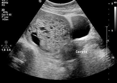
Snowstorm appearance in a complete hydatidiform mole
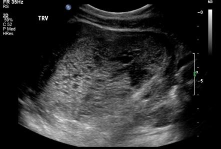
Partial hydatidiform mole