Acute Otitis Media (AOM)
Acute otitis media AKA Acute Suppurative Otitis Media is an acute infection of the middle ear canal. Strictly, it is defined as the first 3 weeks of a process in which the middle ear shows signs and symptoms of acute inflammation. AOM frequently follows an URTI which predisposes to a bacterial infection. Infectious organisms spread to the middle ear through the eustachian tube, external ear if there is traumatic perforation, and blood (uncommon). 60% of AOM cases resolve spontaneously in 24 hours. 80% resolve within 2-3 days. AOM may be initially observed in uncomplicated cases in children > 6 months.
AOM is the second most common disease in children (after URTI). It is the most common infection necessitating treatment for children younger than 5 years old. Some children may get Recurrent AOM, whereby episodes recur up to 5 times a year.
Normal tympanic membrane– pale, pinkish in colour, visualize handle of malleus and cone of light
The tympanic membrane in AOM – slight tympanic membrane bulge, a meniscus of purulent effusion at the bottom of the tympanic membrane, thickened and bulging tympanic membrane
- Risk factors
- Recurrent URTI, Measles, Diphtheria, or whooping cough
- Ages 6-24 months of age – the Eustachian tube is shorter and straighter allowing for easier spread of pathogens.
- Factors contributing to Eustachian Tube Dysfunction
- Craniofacial or skull base abnormalities e.g. Cleft palate
- Recurrent URTI, Measles, Diphtheria, or Whooping Cough
- Allergic Rhinitis
- Nasopharyngeal Tumor
- Chronic Rhinosinusitis
- Tonsilitis and adenoiditis
- Prematurity
- Nasal packing
- Environmental factors
- low SES (poor housing and overcrowding)
- Daycare attendance
- Use of a pacifier
- Passive smoke exposure
- Bottle-feeding while lying on the back
- Smoking
- Ciliary dysfunction
- Prolonged nasotracheal intubation
- Nasogastric tube
- Causative organisms
- Streptococcus pneumoniae (most common)
- Haemophilus influenzae
- Moraxella catarrhalis
- Gram negative bacilli
- GBS in infants
- RSV may precede/predispose to bacterial infection
- Pathophysiology
- ETD causes negative middle ear pressure
- Transudative fluid collects and persists in the middle ear space
- Stagnant fluid = Nidus for Infection, often triggered by an URTI
- Stages of Acute Otitis Media
- Tubal occlusion: Oedema of the eustachian tube causes blockage leading to absorption of air and negative intratympanic pressure
- Presuppuration: Pyogenic bacteria spread to the middle ear and cause inflammatory exudates to appear
- Suppuration: Pus forms in the middle air and to some extent in the mastoid air cells. Tympanic membrane bulges and can rupture.
- Resolution: Tympanic membrane ruptures releasing pus and relieving symptoms
- Complication: Resolution does not take place with high virulence or immunocompromised patients causing the disease to spread
- Signs and symptoms in neonates
- Irritability and crying
- Difficulty to feed
- Fever
- Ear pulling
- Signs and symptoms in older children and adults
- Fever
- Otalgia or ear tugging
- Hearing loss
- Ear fullness
- Tinnitus
- Headache
- Nausea and vomiting
- Otorrhea
- Otoscopic findings
- Bulging and redness of the tympanic membrane
- Non-mobile tympanic membrane
- The malleus and umbo are not seen well
- Perforation of the tympanic membrane (frequently in the posterior or inferior quadrant)
- Opaque serum-like exudate oozing through the entire tympanic membrane
- Investigations
- Pneumatic otoscopy (insufflation of the tympanic membrane): for formal diagnosis
- Immobility
- Type B (flat) curve
- Tympanocentesis: Most accurate test. Indications for tympanocentesis are as follows:
- Neonates < 6 weeks
- Immunocompromised or immunosuppressed patients
- Patients who have had a complications necessitating culture
- Patient with treatment failure.
- Audiogram
- CHL < 30dB
- CT Scan: for complications
- MRI: particularly for intracranial complications
- Pneumatic otoscopy (insufflation of the tympanic membrane): for formal diagnosis
- Medical Treatment
- Oral Antibiotics: Amoxicillin/clavulanate or 2nd/3rd Generation Cephalosporin e.g. Cefuroxime, Ceftriaxone or Macrolide e.g. Azithromycin, Clindamycin
- Standard duration < 6 years: 10 days (except azithromycin which is given for 5 days)
- Standard duration > 6 years and non-severe: 7- 10 days
- Adjunctive Therapy
- Antipyretics and Analgesics e.g. Acetaminophen, NSAIDS
- Decongestants e.g. Oxymetazoline to reduce edema in the middle ear and promote ventilation of the middle ear
- Myringotomy and insertion of a Tympanostomy tube is indicated if there are repeated infections
- Antibiotic prophylaxis for Recurrent AOM
- Full course antibiotics for 10 days
- Reduced bedtime dose for 5-6 weeks
- Oral Antibiotics: Amoxicillin/clavulanate or 2nd/3rd Generation Cephalosporin e.g. Cefuroxime, Ceftriaxone or Macrolide e.g. Azithromycin, Clindamycin
- Indications for Myringotomy and insertion of a Tympanostomy
- Recurrent AOM > 4 episodes in 6 months
- Recurrent AOM > 5-6 episodes in 12 months
- Severe otalgia
- Toxic patients
- Associated complications
- When can antibiotics be delayed in AOM? Low-risk groups. Symptomatically treated for 48-72 hours
- 24 months old
- 6-24 months if the diagnosis is uncertain and the child is clinically well
- What are other indications for antibiotic treatment?
- Temperature on physical exam or by history of > 38.2 degrees Celsius by any method within the past 48 hours
- Symptoms suggestive of AOM for > 48 hours
- Toxic-appearing child
- The tympanic membrane of the infected ear not intact: pus discharging
- The presence of a chronic condition that may impede the child’s immunity or ability to clear the infection, as judged by the clinician
- Another episode of AOM within the past 3 months
- Signs of impending perforation in the infected ear: bulging
- Co-existing bacterial infection
- The family is probably unable to seek medical attention if the child’s clinical status worsens according to the clinician
- The child’s parent or guardian cannot gain an acceptable understanding of the protocol according to the clinician or according to them
- Complications Complications are rare since AOM is a benign condition if treated with antibiotics
- Acute Mastoiditis
- Subperiosteal abscess
- Facial paralysis
- Labyrinthitis
- Petrositis
- Extradural abscess
- Meningitis
- Brain abscess
- Lateral Sinus Thrombophlebitis
- Delayed speech
- Neck abscess

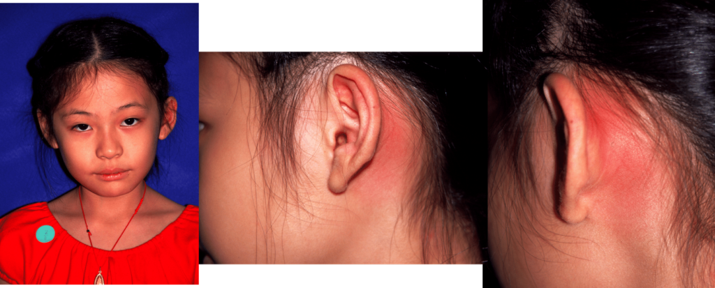
Acute Mastoiditis
Acute mastoiditis is usually seen as a complication of acute otitis media. Symptoms of mastoiditis occur 1-2 weeks after initial onset of acute otitis media, which may or may not have been treated with antibiotics
- Patient History
- H/O acute otitis media (ear pain, fever, vomiting)
- Signs and symptoms
- Pain and tenderness over the mastoid bone
- A red bulging tympanic membrane
- Fever
- “Outstanding ear” from swelling over the post-auricular crease
- Abscess over the mastoid bone
- Appears sick-looking or toxic
- Facial nerve paralysis (rare cases)
- Treatment
- Myringotomy (to drain the middle ear)
- IV antibiotics
- Incision and drainage or aspiration of post-auricular abscess (over the mastoid process)
- Mastoidectomy if the patient remains febrile or toxic or their clinical condition worsens