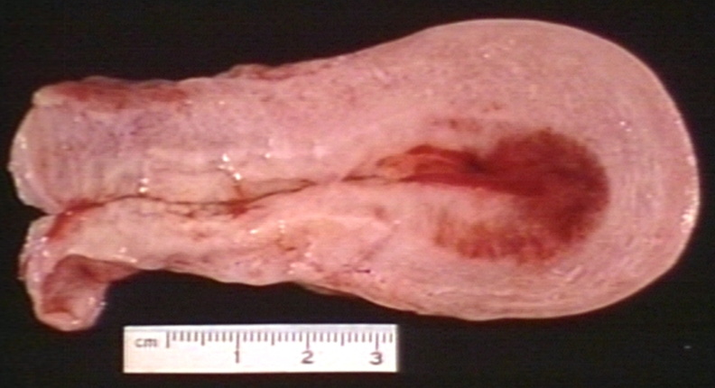
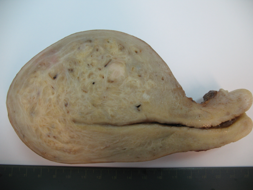
Adenomyosis is uterine enlargement caused by focal or diffuse ectopic rests of endometrial tissue within the myometrium. Pathogenesis is not fully understood. Most likely due to direct invasion of the endometrium into the myometrium. The most common symptoms are abnormal uterine bleeding and secondary dysmenorrhea. Severity of symptoms correlates with foci and extent of invasion. Treatment focuses on controlling pain and bleeding.
Most women with adenomyosis present in their 40s and 50s (older but pre-menopausal – should get better with menopause). Most women with endometriosis present at a younger age. Most of the cases of adenomyosis are diagnosed during hysterectomy (20-60%). No racial differences. Many women with adenomyosis also have fibroids and may be at risk of endometrial cancer (likely have higher estrogen levels)

- Risk factors
- Age: 80% of cases in 40s-50s
- Multiparity: 90% of cases in parous women
- Prior uterine surgery
- Smoking
- Antidepressants (alter the balance of prolactin?)
- **Tamoxifen (**estrogen agonist in the uterus)
- Pathophysiology of menorrhagia
- Increased vascularization of the endometrium near the adenomyotic foci
- Ectopic estrogen production
- Similarities between fibroids and endometriosis
- Characterized by ectopic endometrial tissue
- Both cause pelvic pain
- Both are hormonally snesitive
- Both cause pain and are related to elevated prostaglandin levels (endometrial tissue in both have elevated COX-2)
- Both significantly remit after menopause
- Signs and symptoms
- Abnormal Uterine Bleeding
- Secondary Dysmenorrhea (Pelvic pain)
- Similar to uterine contractions in labour
- May not be localized to the uterus or pelvis (endometrial implants on the uterosacral ligaments may localize/refer to the back)
- Dyspareunia (10%)
- Subfertility (Fertility issues is not as common as with endometriosis – women develop adenomyosis later in life as compared to endometriosis; However there is an increased risk of ectopic pregnancy)
- Physical exam
- Visual inspection: Normal
- Speculum exam: Normal
- Bimanual exam: enlarged and tender uterus
- Diffusely enlarged and boggy in adenomyosis
- Focal adenomyosis can present as fibroids, eliciting a “lumpy, bumpy” texture
- Investigations
- Transvaginal ultrasound: best initial test.
- Increased thickness of the myometrium
- Myometrial heterogeneity
- Small hypoechoic cysts in the myometrium
- Striated projections from endometrium into myometrium
- MRI: more accurate. Used to distinguish adenomyosis from fibroids.
- Other labs
- Urine hCG
- Urinalysis and culture
- Vaginal and cervical cancer
- CBC
- Transvaginal ultrasound: best initial test.
- Treatment
- Combined oral contraceptives or Progestins: shrink foci, endometrial tissue will not be active (reduces prostaglandin production), controls bleeding – first line
- Relieve pain: NSAIDs (for breakthrough pain or for women wishing to become pregnant)
- Surgical treatment
- Hysterectomy: most definitive
- Endometrial ablation (controversial – surgery on the uterus can worsen adenomyosis)
- Uterine artery embolization (controversial)


