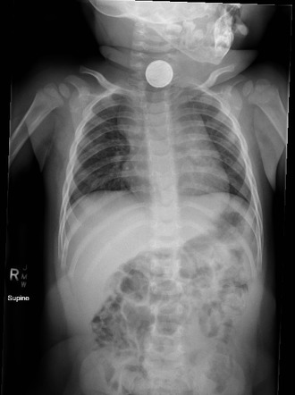
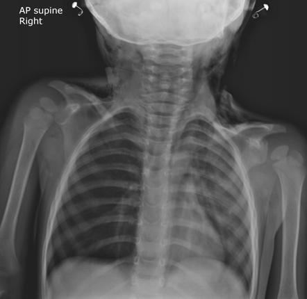
Foreign body of the Esophagus
The most common non-food objects ingested in children are coins. In adults, the most common objects are fish bones, dentures, and meat. The most common locations for impaction are at the cricopharyngeus, gastroesophageal junction, aortic arch, and left mainstem bronchus.
- Signs and symptoms
- Respiratory complaints in children
- Dysphagia
- Drooling
- Weight loss
- Chest pain
- Fever
- Investigations
- X-ray of the Chest, Neck, and Abdomen: to identify radio-opaque objects
- Disk battery: Double-ring shadow on AP and Step-off on Lateral XR
- X-ray of the Chest, Neck, and Abdomen: to identify radio-opaque objects
- Treatment
- Obtain a description and location of the object to aid removal
- Initial observation (for a coin at the cricopharyngeus)
- Rigid esophagoscopy to remove the object
- Refer if:
- Emergent for disk batteries and sharp objects
- For objects causing significant symptoms: complete obstruction or pooling of saliva
- To remove objects not passing within 24 hours
- Children with esophageal anomalies
- Complications
- Esophageal perforation
- Esophageal fistula
- Esophageal stricture
- Mediastinitis
- Pneumomediastinum
- Pneumothorax
- Aspiration

Foreign body of the Airway
The most common foreign bodies in the airway are food (peanuts are the most common) and coins. They are lodged most commonly at the right mainstem bronchus or subsegmental bronchi
- Signs and symptoms
- Violent paroxysms of coughing and choking
- Cough (chronic)
- Wheeze
- Decreased breath sounds
- Stridor
- Investigation
- CXR with inspiratory and expiratory phases: to evaluate for air trapping for non-radiopaque objects
- Air-trapping and Hyperinflation of the affected side (ball-valve effect)
- Hypoinflation of the affected side (atelectasis)
- CXR left/right lateral decubitus phases since the affected lung may not undergo normal collapse when dependent
- Neck X-ray
- CXR with inspiratory and expiratory phases: to evaluate for air trapping for non-radiopaque objects
- Treatment
- Obtain a description of the object and location to aid removal
- Prepare for bronchoscopy
- Decompress stomach with NG Tube to prevent aspiration
- Rigid bronchoscopy under light sedation and spontaneous ventilation.
- Give intra-op Corticosteroids to minimize edema. May require multiple endoscopies
- Post-op Treatment
- Oral Corticosteroid for edema
- Prophylactic Antibiotics
- Close-follow-up
- Complications
- Airway edema
- Death
- Pneumonitis
- Post-obstructive pneumonia
- Pneumothorax
- Chronic lung infection
- Bronchiectasis
Caustic Ingestion
Caustic ingestion causes acute inflammation of the esophagus. The agent ingested can be alkaline or acidic. It may be swallowed accidentally by children, or taken with the purpose of suicide in adults.
The severity of corrosive burns to the esophagus depends on:
- Nature of corrosive substance
- Quantity and concentration of the substance
- Duration of contact with the esophageal wall
Caustic Substances
| Substance | pH | Examples | Pathogenesis |
|---|---|---|---|
| Alkaline Substances | > 11.5 | Laundry detergent, lye, hair straightener, disk battery, drying agents | Causes liquefactive necrosis. Deeper penetration, more severe. Causes saponification of fat, dehydration and thrombosis of blood vessels. More likely to heal with fibrosis. |
| Acidic Substances | < 2 | Battery fluid, toilet cleaner, sulfuric acid, swimming pool cleaners | Causes coagulative necrosis. The coagulum limits penetration |
| Bleach | ~ 11 | Mild irritation, no significant morbidity |
Esophageal burns
| Degree | Extent | Clinical Features |
|---|---|---|
| First Degree | Superficial mucosal injury | Mild hyperemia and edema |
| Second Degree | Transmucosal injury (mucosa, submucosa, possible muscular) | White exudate, erythema |
| Third Degree | Transmural injury (Full-thickness) | Black coagulum, extension beyond the esophagus |
Zarger classification
| Classification | Description |
|---|---|
| 0 | Normal |
| I | Edema and hyperemia of the mucosa |
| IIA | Superficial Ulceration |
| IIB | Deep ulceration |
| IIIA | Focal necrosis |
| IIIB | Extensive necrosis |
| IV | Perforation |
- The three stages of esophageal burns
- Stages of acute necrosis
- Stage of granulations: slough separates leaving a granulating ulcer
- Stage of stricture formation: fibrosis begins after 2 weeks and continues for 2 months or longer
- Signs and symptoms
- Drooling
- Mouth pain
- Stridor (edema of the epiglottis)
- Dysphagia
- Odynophagia
- Refusal to feed in children
- Chest and abdominal pain (does not reliably predict severity)
- General Management
- Call for help, assess vitals, and evaluate the airway (edema may quickly compromise the airway)
- A- Airway opening maneuvers, if there is persistent stridor consider a tracheostomy
- B – Check RR, SpO2 and Give oxygen as needed
- C- Check HR, BP, Peripheral pulses, Cap Refill. Fix IV lines and obtain samples. Give IV fluids as needed.
- D – GCS/AVPU in child, Pupils, and Motor and sensory assessment, Measure RBS
- E – Expose the patient and look for other injuries
- Obtain an AMPLE History
- Determine the type of caustic ingested and when it was ingested
- Rule out signs and symptoms of shock, upper airway obstruction, mediastinitis, peritonitis, acid-base imbalance, and associated burns of the face, lips, and oral cavity
- Have the patient drink water or irrigate the mouth with water. DO NOT LAVAGE or INDUCE EMESIS ****(this will give the substance a “second pass” through the anatomy to cause more damage)
- Identify the agent, and determine pH if necessary
- Direct Esophagoscopy at 24-48 hours (ideally) to evaluate for injury
- Perform too early (<12 hours) and the severity of damage may be missed
- Performed too late (> 48 hours) and may risk perforating the esophagus
- Barium swallow Esophagogram to evaluate for stricture formation or to confirm perforation
- Should be the first diagnostic tool instead of Esophagoscopy if the patient arrives > 48 hours
- Prevent stricture formation: IV corticosteroids- 0.1mg/ kg/ dose TDS within 48- 96 hours for 4- 6 weeks
- Analgesia
- PPI/ Antacids
- Prophylactic antibiotics: IV/ IM for 3-6 weeks depending on the degree of the burns
- NG tube for feeding. Give water or milk, also maintains esophageal lumen
- NPO for 7- 10 days
- Serial esophagograms/ barium swallows every 2 weeks till healing is complete
- Baseline investigations: CBC, arterial blood gases, UECs, CXR, Random Blood Sugar
- Chest-Xray for mediastinitis
- Abdominal series for Gastric perforation
- Arterial Blood Gas for acid-base disturbance
- Treatment of first-degree esophageal burns
- Immediate
- Irrigation
- Observation
- Long-term
- Anti-reflux agents
- Follow-up 2 weeks and consider esophagram if symptomatic
- Immediate
- Treatment of second-degree esophageal burns
- Immediate
- Irrigation
- Observe overnight
- Consider Nasogastric tube or Silastic stent if circumferential
- Long-term
- Corticosteroids
- Antireflux agents
- Antibiotics
- Esophagram to evaluate for strictures
- Immediate
- Treatment of third-degree esophageal burns
- Immediate
- Secure airway (may require tracheotomy)
- Esophagectomy/Gastrectomy with Exploratory laparotomy to remove necrotic tissue
- Consider NG tube or Silastic stent for minimal necrosis
- Monitor overnight
- Second-look at 24 hours
- Long-term
- Broad spectrum antibiotics
- Reflux regimen
- Lathyrogenic agents to prevent stenosis
- Follow-up esophagram at 1 month then q3months x 4 to evaluate for structures
- Immediate
- Stricture management methods:
- Esophagoscopy and prograde dilatations, if permeable
- Gastrostomy and retrograde dilatation, if impermeable
- Esophageal reconstruction or by-pass, if dilatations are impossible
- Why are corticosteroids contraindicated in third-degree esophageal burns?
- May mask infection
- May increase the risk of perforation
- Worsens healing
- Complications
- Stricture formation (circumferential burns)
- Pneumonia
- Tracheoesophageal fistula
- Laryngeal edema
- Perforation
- Hemorrhage and Shock
- Mediastinitis
- Peritonitis
- Esophageal carcinoma (caustic ingestion increases risk 1000x)
- Death