Overview
- Classification of anaerobes
- Obligate anerobes
- Facultative anerobes
- Aerotolerant anerobes.
- Characteristcs and isolation of anaerobes
- Growth of anerobes is inhibited by oxygen due to reduced amount of catalase and SOD.
- Areas of Low Eh (oxidation-reduction potential) support their growth (periodontal pocket, dental plaque, colon, crushing injuries that impair blood supply).
- Habitats include the environment, contaminated foods and GIT of animals.
- In humans, they are found in the oral cavity, GIT, lower GUT and skin.
- Specimen are collected as far as possible from their normal resident sites and are not allowed to dry (dessication reduces the number of bacteria)
- Effective cleaning to avoid contamination using needle + syringes since swabs may dry out.
- Stuart’s transport media is used in swab collected specimen.
- Transport is via an oxygen free system (Syringe + needle, tube or vial or sterile screw cap container).
- Inoculate into specific media promptly and incubate in anerobic conditions (McIntosh-Filde’s Jar, Brewer anaerobic Jar, Aanerobic Glove box, Candle jar).
- Microscopic exam is via gram stain, dark field or phase contrast (spirochetes), Fluorescent Ag staining (Bacteroides).
- Gas liquid chromatography is done on exudates and body fluids to demonstrate products of metabolism (butyric acid, oxybutryic acid, proprionic acid).
- Anerobic culture methods include non-selective media, enrichment media, selective media (BA with Neomycin), Liquid media (Robertson Cooked-Meat Media, Thioglycollate, Blood Culture Media).
- Anerobic media include Roberston cooked media and Thioglycolate media.
- For the environment, air evacuation and replacement with inert gases (H2, N2, CO2),
- Use of disposable gas generation kits (release H2, CO2, N2) anaerobic jars.
- Metyhlene blue is used as an indicator in Brewer or anaerobic jar. Palladium catalyses the reaction causing H20 and condensation.
- Quality control is via chemical indicators (Methylene blue) that change colour when oxygen is removed or viable Biological organism that grow only in the presence of O2 or absence of CO2.
- Antimicrobial susceptibility to metronidazole and clindamycin.
- More resistant to aminoglycosides (Requires O2 to enter cells).
- Anaerobes according to their morphological characteristics
- Gram positive
- Cocci: Peptostreptococcus, Peptococcus
- Spore forming bacilli: Clostridium spp.
- Non-spore forming bacilli: Actinomyces spp., Bifidobacterium spp. , Propionibacterium spp., Lactobacillus spp. ,Mobiluncus spp.
- Gram Negative
- Cocci: Veillonella
- **Non-spore forming bacilli: **Bacteroides spp. , Fusobacterium spp. , Prevotella spp. , Porphyromonas spp.
- Spirochetes: Treponema spp, Borrelia spp
- Gram positive
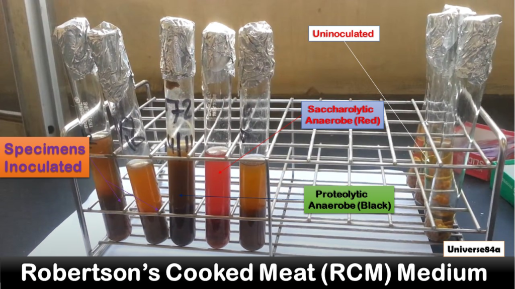
- Components and interpretation of Robertson’s cooked meat media (RCM)
- RCM Contains nutrient broth ,and minced and cooked ox-heart tissue. Unsaturated fatty acids in minced meat act as a reducing agent. Liquid paraffin in layered over the broth to prevent entry of air. Reactions:
- Proteolytic reaction: Blackening of the meat, very unpleasant smell due to protein decomposition, Clostridium tetani
- Saccharolyic reaction: Reddening of meat, rancid smell due to CHO decomposition. Saccharolytic anerobes: Clostroidium perfringens
- RCM Contains nutrient broth ,and minced and cooked ox-heart tissue. Unsaturated fatty acids in minced meat act as a reducing agent. Liquid paraffin in layered over the broth to prevent entry of air. Reactions:
- Gram negative anerobic bacilli
- There are more than 30 genera of anerobic GNRs.
- They are found in the mouth, GIT, vagina and are among the most important constituents of normal flora.
- They cause a variety of infections in humans, particularly polymicrobial infecitons and abscsses.
- Human infections are largerly restricted to:
- Bacteroides spp.
- Prevotella spp.
- Porphyromonas spp.
- Fusobacterium spp.
Bacteroides
- Bacteroides
- Bacteroides is a pleomorphic GN bacillus or coccobacillus that is non-spore forming, non-motile, and encapsulated.
- It is an anerobic rod that leads to opportunistic and polymicrobial infections.
- The species of medical importance is Bacteroides fragilis, which is a predominant commensal of the colon (approximate numbers 10^11/g of feces) and vagina (60% of femals)
- Bacteriodes corrodens is found in the oral cavity.
- Conditions predisposing to infection with Bacteroides fragilis
- surgery
- trauma
- chronic diseases
- Factors contributing to infection with Bacteroides fragilis
- Local tissue necrosis
- Impaired blood supply
- Growth of facultative anerobes at the site (facultative anerobes {such as E.coli} utilize oxygen, reducing it to a level that allow anaerobes {Bacteroides and Prevotella} to thrive)
- Virulence factors associated with Bacteroides fragilis
- Capsular polysaccharide: inhibits opsonization/ phagocytosis, promotes abscess formation, promotes adherence to epithelial cells
- Pili and fibriae: adherence to epithelial cells and mucus
- Succinic acid: inhibits phagocytosis and intracellular killing
- LPS endotoxin: less toxic compared to other GN bacteria (infections do not directly produce signs of sepsis that common in other GN bactera)
- Enzymes (heparinase, fibrinolysins, hyaluronidase, neuraminidase): tissue damage, promotes invasion and spread
- Catalse and SOD: tolerates oxygen exposure mitigating toxic effects of oxygen and facilitating survival and propagation of the organism
- Heat-labile Zn metalloproteinase toxin (B.fragilis toxin): In enterotoxigenic strains, causes morphological changes on the intestinal epithelium via F-actin rearrangement (causing chloride secretion and fluid loss), Also induces IL-8 secretion (contributes to inflammatory damage)
- Clinical features of Bacteroides fragilis
- Intra-abdominal infection
- Abscesses: common anaerobic isolate (polymicrobial), foul smell of infections or discharge due to metabolism of Short Fatty acid chains (succinic and butyric acid)
- Diarrhea: Enterotoxin producing strains in children
- Bacteremia: commonest isolate in anerobic bacteremia, source is usually intra-abdominal and is associated with abscesses, malignancy, bowel perforation or surgery. Septic shock is less common in B.fragilis than in bacteremia caused by aerobic GNRs due to absence of lipid A in the endotoxin of B.fragilis
- Endocarditis: large vegetations and high frequency of thromboembolic complications
- Skin and soft tissue infections: part of mixed flora in diabetic and decubitus ulcers
- Bone and joint infections: rare causes of osteomyelitis and septic arthritis
- CNS infection: anaerobic meningitis is rare, though it is the commonest isolate. Anaerobes are frequently implicated in brain abscesses.
- Head and neck disease
- Intra-abdominal infection
- Laboratory features of Bacteroides fragilis
- Specimen: Pus, exudate, infected tissue, blood (Specimen suspected or containing anerobes must be delivered to the lab ASAP and cultured anerobically with minimum delay)
- Culture
- BA: Selective/ enriched by adding kanamycin and vancomycin;
- grey, glistening, non-hemolytic, 1-2 mm diameter colonies (usually within 48H of anerobic incubation)
- Stringy colonies (when emulsified in3% KOH, form long strands when the loop is pulled away, remember the cholera string test?)
- BA: Selective/ enriched by adding kanamycin and vancomycin;
- Gram stain: Pale pink, pleomorphic coccobaclli with irregular or bipolar staining
- Growth in 20% bile: differentiates from other anerobic GNRs
- Gas Liquid Chromatography: Biochemical reactions (Suger fermentations) and byproducts of certain organic acids (formic, acetic and propionic acid
Prevotella and Porphyromonas
- Prevotella
- Morpholgy is like that of Bacteroides.
- Species include:
- Prevotella melaninogenica
- Prevotella bivia
- Prevotella oralis
- Prevotella buccalis.
- Are normal flora of the oral cavity.
- Involved in mixed infections:
- URTI
- Abscesses (mouth, brain and lung)
- Pelvic and abdominal infections.
- Porphyromonas
- Still similar to Bacteroides.
- Porphyromonas are non-spore forming, non-motile, anaerobic, GN bacillus.
- Species include:
- Porphyromona gingivalis
- Porphyromonas endodontalis
- Porphyromonas asaccharolytica.
- They are normal flora of the mouth.
- Causes gingival and tooth infection and various abscesses.
- Laboratory features of Prevotella and Porphyromonas
- Non-spore forming, non-motile, anerobic gram negative bacilli
- Usually isolated (along with toher anerobes) from abscesses and soft tissue infections
- May both appear pigmented (usually brown/black)
- Young unpigmented colonies show brick-red fluorescence under UV lighe (Prevotella melaninogencia)
- Growth is inhibited by 20% bile
- Prevotella – moderately saccharolytic
- Porphyromona – asaccharolytic
Fusobacterium
- Fusobacterium
- Fusobacterium is part of mixed infections with facultative anerobes or strict anerobes from intra-abdominal abscesses.
- Isolated single from blood culture on skin or mucus membrane/RT infections occasionally.
- Fusobacterium necrphorum causes severe systemic infections (Lemierre’ disease – in previously healthy young adults, severe sore throiat, fever, cervical lymphadenopathy and unilateral thrombophlebitis of the internal jugular)
- Fusobacterium nucleatum causes ulcerative gingivitis (Vincent’s angina – also Borellia vincenti).
- They are susceptile to penicillin, metronidazole and clindamycin.